Cardiac Conduction System Diagram
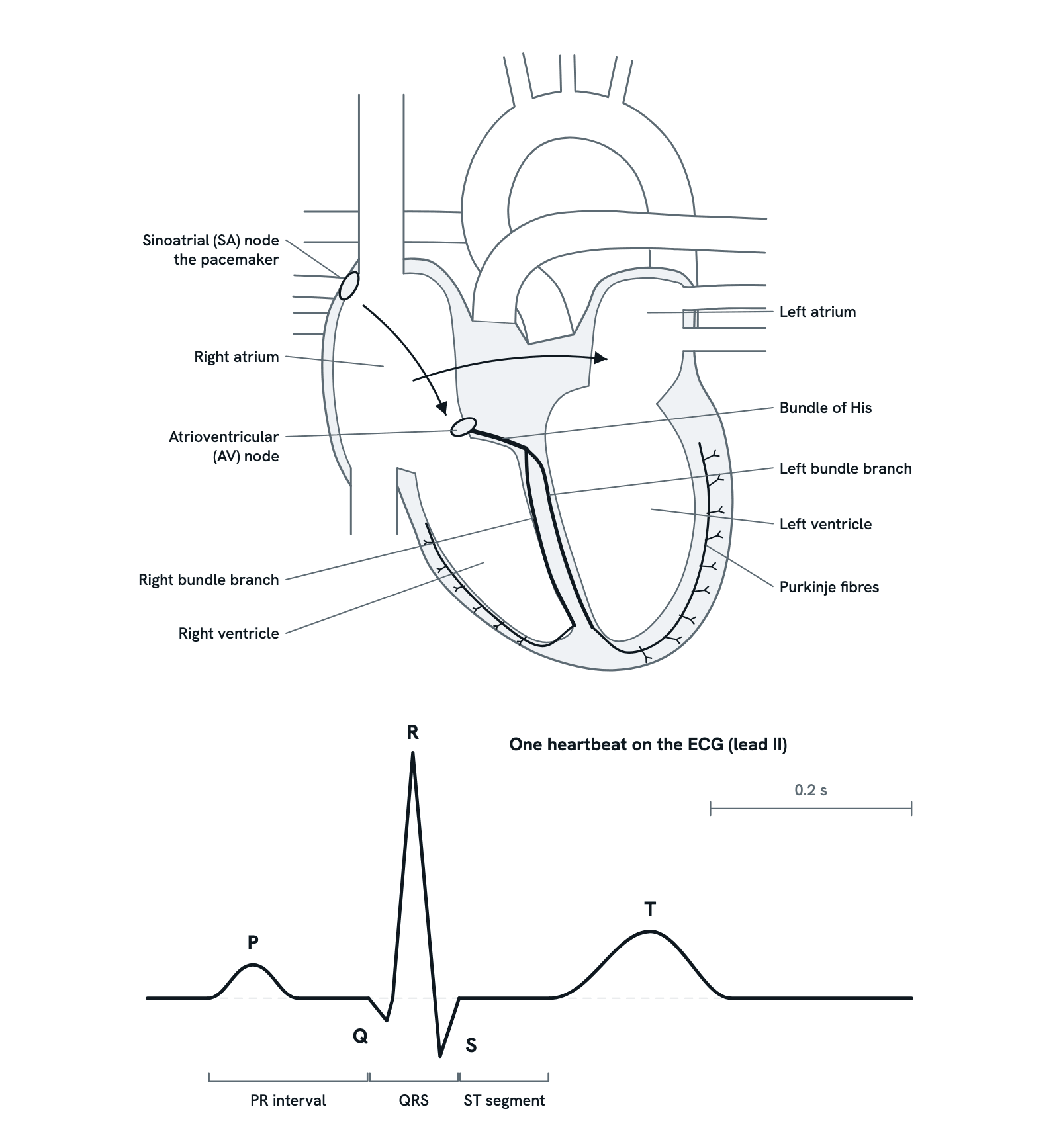
How the heart's electrical impulse travels from the SA node to the Purkinje fibres, and the stage of the beat behind each ECG wave: P, QRS and T.
Each heartbeat is set off by an electrical impulse the heart makes itself. It starts in the sinoatrial node, spreads across the atria, is held back at the atrioventricular node, then races through the ventricles. Under the heart is one beat on an ECG, each wave matched to its stage. A simplified section seen from the front; not to scale. Timings are typical for a resting adult.
Also called Electrical conduction system of the heart or Conduction system of the heart.
Step 1 of 9
The heartbeat's electrical path
The heart makes its own electrical impulse at every beat and carries it along a set path of specialised cells: the SA node, the atria, the AV node, the bundle of His, the bundle branches and the Purkinje fibres. An ECG records the whole beat from the skin, and each wave matches a stage of the journey.
Use ← and → to move between steps.
Step 1 of 9: The heartbeat's electrical path
Download the diagramChecked against its sources on 29 October 2026.
Step by step
The steps of the Cardiac Conduction System
01The heartbeat's electrical path
The heart makes its own electrical impulse at every beat and carries it along a set path of specialised cells: the SA node, the atria, the AV node, the bundle of His, the bundle branches and the Purkinje fibres. An ECG records the whole beat from the skin, and each wave matches a stage of the journey.
02The SA node fires
The sinoatrial node, a small cluster of pacemaker cells in the wall of the right atrium near the superior vena cava, fires on its own. At rest it usually sets a rate of about 60 to 100 beats a minute, which the autonomic nerves speed up or slow down. Its firing is too small to see on the ECG.
03Across the atria: the P wave
The impulse spreads from cell to cell through the muscle of both atria, reaching the left atrium through Bachmann's bundle. As the atria depolarise, the ECG records the small, rounded P wave, and the atria then contract, pushing the last of their blood into the ventricles.
04Held back at the AV node: the PR interval
The impulse reaches the atrioventricular node, the only normal path from the atria to the ventricles, and slows down there. The pause lets the atria finish emptying before the ventricles contract. On the ECG the line stays flat after the P wave; the whole PR interval, from the start of the P wave to the start of the QRS, is normally about 0.12 to 0.20 s.
05Into the bundle of His and the bundle branches
From the AV node the impulse passes into the bundle of His, which crosses the insulating tissue into the top of the interventricular septum and divides into the right and left bundle branches, one down each side of the septum. These fibres are too small to show on the ECG, so their part falls within the PR interval. The septal muscle is activated first, which can give a small first dip, the Q wave, at the start of the QRS complex.
06Through the Purkinje fibres: the QRS complex
The bundle branches end in Purkinje fibres, which carry the impulse very fast through the walls of both ventricles, so all their muscle is activated almost at once. As it depolarises, the ECG records the tall, narrow QRS complex, normally under about 0.12 s. The atria repolarise at about the same time, but their wave is very small and falls mostly within the QRS, so it is not normally seen.
07The ventricles contract: the ST segment
With all the ventricular muscle depolarised, the trace returns to the baseline and stays flat: the ST segment. This is while the ventricles contract and push blood into the arteries. A raised or lowered ST segment can be a sign of heart muscle short of oxygen or damaged, as in a heart attack.
08The ventricles recover: the T wave
The ventricular muscle repolarises, ready to be activated again, and the ECG records the broad, rounded T wave. The ventricles then relax and fill again before the next beat.
09Until the next beat
After the T wave the trace stays flat until the SA node fires again and the next P wave begins. At a resting rate of about 75 beats a minute, one whole beat, from P wave to P wave, takes about 0.8 s.
Parts
What each part of the cardiac conduction system does
- Sinoatrial (SA) node
- A small cluster of pacemaker cells in the wall of the right atrium, where the superior vena cava joins it. Its cells fire on their own, faster than any other part of the heart, so it sets the heart rate.
- Atria
- The impulse spreads from cell to cell through the muscle of both atria, and on to the left atrium through a band called Bachmann's bundle. As the atria depolarise they make the P wave, and then they contract.
- Atrioventricular (AV) node
- Low in the right atrium, on the septum between the atria. Fibrous tissue insulates the atria from the ventricles everywhere else, so this is the only normal way through, and it conducts slowly, holding the impulse back for a fraction of a second.
- Bundle of His
- A short bundle of conducting fibres that carries the impulse from the AV node through the insulating tissue into the top of the interventricular septum, where it divides.
- Bundle branches
- The right and left bundle branches run down either side of the interventricular septum towards the apex. If one is blocked, that ventricle is activated late and the QRS complex widens.
- Purkinje fibres
- Fast-conducting fibres that branch out from the bundle branches through the walls of both ventricles, so that all of the ventricular muscle is activated almost at once.
- Ventricles
- The Purkinje fibres activate their muscle almost all at once, so both ventricles contract together in one coordinated squeeze. Their depolarisation makes the QRS complex and their repolarisation the T wave.
- P wave
- A small, rounded wave made as the atria depolarise. It does not show the SA node firing, which is too small to record from the skin.
- PR interval
- From the start of the P wave to the start of the QRS complex, normally about 0.12 to 0.20 s. It includes the delay in the AV node; a longer interval means the impulse is held up on its way to the ventricles, most often in the AV node, as in first-degree heart block.
- QRS complex
- The tall, narrow spike made as the ventricles depolarise, with a downward Q, a tall R and a downward S. It normally lasts under about 0.12 s, usually 0.06 to 0.10 s; a wider QRS means slowed spread through the ventricles.
- ST segment
- The flat stretch between the QRS complex and the T wave, while all the ventricular muscle is depolarised and contracting. It normally lies level with the PR segment; raised or lowered, it can be a sign of heart muscle short of oxygen or damaged.
- T wave
- A broad, rounded wave made as the ventricles repolarise, recovering so that they can be activated again.
Dictionary
The terms to know
Practise it
The quizzes and cards that fit
- Physiology quizzes
- Exercise Physiology15 questions
- Puberty15 questions
- Blood Clotting15 questions
- Medicine quizzes
- Labor and Delivery15 questions
- IV Fluids15 questions
- Sepsis15 questions
- Physiology flashcards
The subject's terms as a ready-made deck, free to flip through.
Next to it
More diagrams
Sources
Where the facts come from
- StatPearls: Physiology, Cardiac (NCBI Bookshelf)
- Becker, Fundamentals of Electrocardiography Interpretation, Anesthesia Progress, 2006 (PMC)
- NHLBI: How the Heart Beats
- NHLBI: Conduction Disorders
- StatPearls: Anatomy, Thorax, Heart (NCBI Bookshelf)
- StatPearls: Electrocardiogram (NCBI Bookshelf)
- StatPearls: P Wave (NCBI Bookshelf)
- StatPearls: Physiology, Cardiac Cycle (NCBI Bookshelf)
- Manne, Atrial Repolarization Waves (Ta) Mimicking Inferior Wall ST Segment Elevation Myocardial Infarction, Case Reports in Medicine, 2018 (PMC)
Drawn and written by biofideX from these sources.