Human Heart Diagram
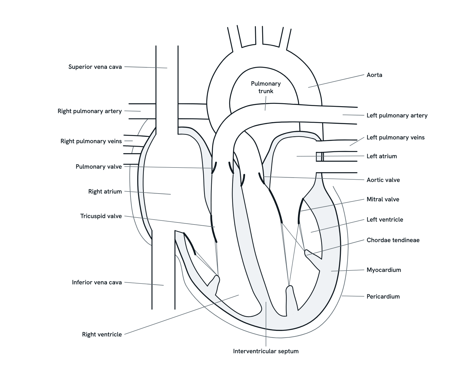
The parts of the human heart and what each does: the four chambers, the four valves, the septum, the wall and the great vessels that carry blood in and out.
The heart is a muscular pump with four chambers: two atria that receive blood and two ventricles that pump it out, the right side to the lungs and the left side to the rest of the body. This is a section seen from the front, with the patient's right on your left, as textbooks draw it. Not to scale. Press any part, or take the tour.
Also called Heart.
Press any part of the diagram, or pick one here, to see what it is and what it does.
Download the diagramChecked against its sources on 29 October 2026.
A tour
A tour of the human heart
01The heart at a glance
The heart is two pumps side by side. The right side sends oxygen-poor blood to the lungs; the left side sends oxygen-rich blood to the rest of the body. Each side has an atrium that receives blood and a ventricle that pumps it out, and four valves keep the blood moving one way.
02The atria receive blood
The two upper chambers, the atria, have thin walls. The right atrium receives oxygen-poor blood from the body through the venae cavae; the left atrium receives oxygen-rich blood from the lungs through the pulmonary veins. Each passes its blood down into the ventricle below.
03The ventricles pump
The two lower chambers, the ventricles, do the pumping. The right ventricle sends blood only to the lungs, at low pressure. The left ventricle sends it round the whole body at a much higher pressure, and its wall is the thickest in the heart. The interventricular septum lies between them.
04The tricuspid and mitral valves
Between each atrium and its ventricle is an atrioventricular valve: the tricuspid on the right, with three cusps, and the mitral on the left, with two. When the ventricles contract, the rising pressure pushes the cusps shut, and the chordae tendineae, pulled tight by the papillary muscles, stop them turning back into the atria.
05The pulmonary and aortic valves
Where each ventricle empties into its artery is a semilunar valve, named for its half-moon cusps. The pulmonary and aortic valves open as the ventricles contract and close as they relax, so the blood cannot fall back. The closing of the valves, first the atrioventricular pair and then these two, makes the two sounds of each heartbeat.
06The great veins bring blood in
The superior vena cava drains the head, neck, arms and chest, and the inferior vena cava the legs, abdomen and pelvis; both empty into the right atrium, as does the coronary sinus, which drains the heart's own muscle. The pulmonary veins, usually two from each lung, carry oxygen-rich blood into the left atrium.
07The great arteries carry blood out
The pulmonary trunk leaves the right ventricle and divides into the right and left pulmonary arteries, one for each lung. The aorta leaves the left ventricle beside it, rises, arches back and to the left, and runs down through the chest; three large arteries for the head and arms leave the arch.
08The wall and the pericardium
The heart's wall has three layers: the thin endocardium lining the chambers and valves, the thick muscle of the myocardium, and the epicardium outside. Round the heart, the pericardium forms a sac; a little fluid between its layers lets the heart beat with little friction.
09The right side: to the lungs and back
The right atrium and ventricle pump oxygen-poor blood through the pulmonary arteries to the lungs, where it gives up carbon dioxide and takes up oxygen in the capillaries round the alveoli. The pulmonary veins bring it back to the left atrium. This loop is the pulmonary circuit.
10The left side: round the body
The left atrium and ventricle pump oxygen-rich blood into the aorta and on through the arteries to the capillaries of every tissue. Veins bring the blood back, now oxygen-poor, to the right atrium. This is the systemic circuit; the two circuits run one after the other, and both sides of the heart beat together.
Parts
What each part of the human heart does
- Right atrium
- Receives oxygen-poor blood from the body through the superior and inferior venae cavae, and from the heart's own veins through the coronary sinus. It passes the blood through the tricuspid valve to the right ventricle.
- Right ventricle
- Pumps oxygen-poor blood through the pulmonary valve into the pulmonary trunk and on to the lungs. The lungs' blood vessels are at low pressure, so it pumps at a much lower pressure than the left ventricle and its wall is thinner.
- Left atrium
- Receives oxygen-rich blood from the lungs through the pulmonary veins and passes it through the mitral valve to the left ventricle.
- Left ventricle
- Pumps oxygen-rich blood through the aortic valve into the aorta and on round the whole body. It works at the highest pressure of the four chambers, so its wall is the thickest.
- Interventricular septum
- The wall of muscle between the two ventricles, which keeps the oxygen-poor blood of the right side apart from the oxygen-rich blood of the left. The interatrial septum separates the two atria in the same way.
- Myocardium
- The heart muscle, the thick middle layer of the wall, whose cells contract together to pump. It is lined inside by the thin endocardium and covered outside by the epicardium, the inner layer of the pericardium.
- Pericardium
- A tough sac round the heart and the roots of the great vessels, which holds the heart in place and protects it. Its thin inner layer folds back over the heart as the epicardium, and a little fluid between the two lets the heart move as it beats.
- Tricuspid valve
- Between the right atrium and the right ventricle. Its three cusps open as the ventricle fills and are pushed shut when it contracts, so blood cannot flow back into the atrium.
- Pulmonary valve
- At the exit of the right ventricle, with half-moon (semilunar) cusps. It opens as the ventricle pumps blood into the pulmonary trunk and closes as the ventricle relaxes, so the blood cannot flow back.
- Mitral valve
- Between the left atrium and the left ventricle; also called the bicuspid valve, because it has two cusps. It shuts when the left ventricle contracts, so blood is not pushed back towards the lungs.
- Aortic valve
- At the exit of the left ventricle, usually with three half-moon (semilunar) cusps. It opens as the ventricle pumps blood into the aorta and closes as the ventricle relaxes. The coronary arteries, which feed the heart muscle, arise just above it.
- Chordae tendineae and papillary muscles
- Fine cords run from the cusps of the tricuspid and mitral valves to papillary muscles on the ventricle walls. The rising pressure in the contracting ventricles shuts the cusps; the papillary muscles contract too and keep the cords taut, so the cusps are not pushed back into the atria.
- Superior vena cava
- Brings oxygen-poor blood from the head, neck, arms and chest into the top of the right atrium.
- Inferior vena cava
- Brings oxygen-poor blood from the legs and from the organs of the abdomen and pelvis into the lower part of the right atrium.
- Pulmonary trunk
- The short, wide artery that leaves the right ventricle through the pulmonary valve, beside the ascending aorta. It soon divides into the right and left pulmonary arteries.
- Pulmonary arteries
- Carry oxygen-poor blood from the pulmonary trunk to the lungs, one to each lung, where they branch again and again down to the capillaries round the air sacs.
- Pulmonary veins
- Usually four, two from each lung, bringing oxygen-rich blood back to the left atrium.
- Aorta
- The body's largest artery. It rises from the left ventricle, arches back and to the left, and runs down through the chest and abdomen. Three branches leave the arch for the head and arms: the brachiocephalic trunk and the left common carotid and left subclavian arteries.
Dictionary
The terms to know
Practise it
The quizzes and cards that fit
- Human Anatomy quizzes
- Shoulder and Rotator Cuff15 questions
- Knee Joint15 questions
- Arteries and Veins15 questions
- Physiology quizzes
- Exercise Physiology15 questions
- Puberty15 questions
- Blood Clotting15 questions
- Medicine quizzes
- Labor and Delivery15 questions
- IV Fluids15 questions
- Sepsis15 questions
- Human Anatomy flashcards
The subject's terms as a ready-made deck, free to flip through.
Next to it
More diagrams
Sources
Where the facts come from
- StatPearls: Anatomy, Thorax, Heart (NCBI Bookshelf)
- NHLBI: How the Heart Works, What the Heart Looks Like
- NHLBI: How Blood Flows Through the Heart
- NCI SEER Training Modules: Structure of the Heart
- NCI SEER Training Modules: Circulatory Pathways
- StatPearls: Anatomy, Thorax, Heart Great Vessels (NCBI Bookshelf)
- StatPearls: Anatomy, Thorax, Heart Papillary Muscles (NCBI Bookshelf)
- StatPearls: Anatomy, Thorax, Pericardium (NCBI Bookshelf)
- StatPearls: Physiology, Cardiac Cycle (NCBI Bookshelf)
- Dal-Bianco, Basic Mechanisms of Mitral Regurgitation, Canadian Journal of Cardiology, 2014 (PMC)
- Bauer, Cardiovascular Anatomy and Pharmacology, in Basic Sciences in Anesthesia, 2017 (PMC)
Drawn and written by biofideX from these sources.