Respiratory System Diagram
The parts of the respiratory system and what each does, from the nose and trachea to the bronchi, the lobes of the lungs and alveoli.
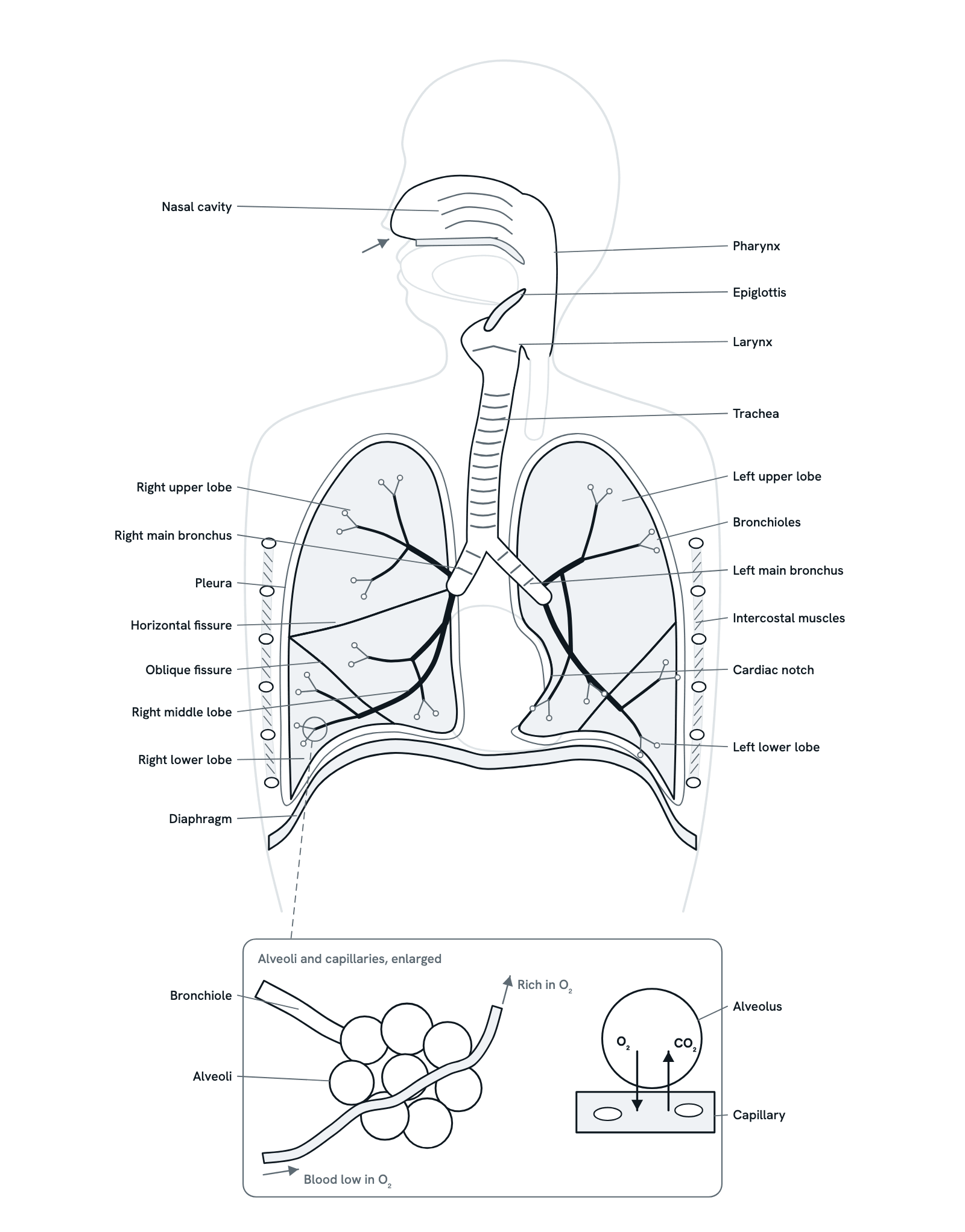
Air reaches the lungs through a branching set of tubes, and oxygen passes into the blood across the thin walls of the alveoli. Press any part to see what it does, or follow a breath in order. Front view of the chest, with the head turned to show the nose and throat in section; the inset enlarges the alveoli. Not to scale.
Also called Human respiratory system or Lungs and airways.
Press any part of the diagram, or pick one here, to see what it is and what it does.
Download the diagramChecked against its sources on 29 October 2026.
A tour
A tour of the respiratory system
01The respiratory system at a glance
Air passes through the nose, pharynx and larynx into the trachea, then down a branching tree of bronchi and bronchioles to the alveoli deep in the lungs. There oxygen moves into the blood and carbon dioxide moves out. The diaphragm and the muscles between the ribs move the air in and out.
02Warming, filtering and moistening
Air comes in through the nose or the mouth. In the nasal cavity, hairs trap large particles, and the moist, mucus-lined walls warm and humidify the air and catch dust, since cold, dry air can irritate the lungs. The air then passes down through the pharynx.
03The larynx and epiglottis
The larynx, or voice box, carries air between the pharynx and the trachea, and its vocal cords vibrate to make sound as air passes out between them. When you swallow, the epiglottis folds down over its opening, so food and drink go into the esophagus instead.
04The trachea
The trachea runs down the neck into the chest. C-shaped rings of cartilage stop it from collapsing, their open backs facing the esophagus. Mucus on its lining traps dust and microbes, and tiny cilia sweep the mucus up to the throat, where it is swallowed or coughed out.
05The airway tree
In the chest the trachea divides into the right and left main bronchi. Each splits into lobar bronchi, one per lobe, and on into smaller and smaller branches, the bronchioles. As the airways narrow, cartilage thins out and smooth muscle takes its place, until the bronchioles have no cartilage at all.
06Lobes and fissures
The right lung has three lobes, separated by the horizontal and oblique fissures. The left lung has two, separated by an oblique fissure, and a cardiac notch where the heart sits, so it is a little smaller. Each lobe has its own lobar bronchus.
07The pleura
Each lung is wrapped in pleura: one layer on the lung's surface, another lining the inside of the chest. The thin film of fluid between them lets the lungs glide against the chest wall as they fill and empty, and holds the two layers together.
08Alveoli and gas exchange
The bronchioles end in clusters of alveoli, tiny air sacs wrapped in capillaries. Their walls are so thin that gases cross them by diffusion: oxygen moves from the air into the blood, and carbon dioxide moves from the blood into the air, to be breathed out.
09Breathing in
Breathing in takes muscle work. The diaphragm contracts and flattens, and the outer intercostal muscles lift the ribs up and out. The chest grows larger, the pressure inside the lungs falls below the pressure of the air outside, and air flows in.
10Breathing out
At rest, breathing out is passive: the diaphragm and intercostal muscles relax, and the stretched lungs and chest wall spring back. The chest grows smaller, the pressure in the lungs rises above the air outside, and air flows out. When you breathe hard, the abdominal muscles help push it out.
Parts
What each part of the respiratory system does
- Nasal cavity
- The air space behind the nose, above the palate. Hairs at its entrance trap large particles, and its moist, mucus-lined walls warm and humidify the air and catch dust before it goes deeper.
- Pharynx
- The throat, a passage shared by air and food. Air from the nose passes down through it into the larynx; food from the mouth passes on into the esophagus behind.
- Epiglottis
- A flap of tissue at the top of the larynx. When you swallow, it folds down over the opening of the larynx, so food and drink go into the esophagus rather than the airway.
- Larynx
- The voice box, between the pharynx and the trachea, built of cartilages joined by muscles and ligaments; the thyroid cartilage forms the Adam's apple. Its vocal cords vibrate as air passes out between them.
- Trachea
- The windpipe. C-shaped rings of cartilage hold it open; their open backs face the esophagus. Its lining makes mucus that traps particles, and cilia sweep the mucus up to the throat.
- Bronchi
- The trachea divides into the right and left main bronchi. The right is wider, shorter and more upright, so inhaled objects lodge on the right more often. Each divides into lobar bronchi, one for each lobe.
- Bronchioles
- The narrow airways the bronchi branch into, over many generations. Unlike the bronchi, they have no cartilage; smooth muscle in their walls can widen or narrow them. The last ones open into the alveoli.
- Right upper lobe
- The top lobe of the right lung, above the horizontal fissure. Its apex rises a little above the first rib, into the root of the neck.
- Right middle lobe
- A wedge at the front of the right lung, between the horizontal and oblique fissures. The left lung has no middle lobe.
- Right lower lobe
- The lowest lobe of the right lung, below the oblique fissure, its base resting on the dome of the diaphragm. Seen from the front, only a small part of it shows.
- Left upper lobe
- The upper of the left lung's two lobes, above the oblique fissure. Its front edge holds the cardiac notch, with a tongue-shaped part, the lingula, below it.
- Left lower lobe
- The lower of the left lung's two lobes, below the oblique fissure, its base resting on the diaphragm.
- Horizontal fissure
- A cleft found only in the right lung, separating the upper lobe from the middle lobe.
- Oblique fissures
- A slanting cleft in each lung. In the right lung it separates the middle and lower lobes; in the left lung, the upper and lower lobes.
- Cardiac notch
- A hollow in the front edge of the left lung that makes room for the heart. It is one reason the left lung is a little smaller than the right.
- Pleura
- Two thin layers round each lung: one covers the lung, the other lines the inside of the chest. A film of fluid between them lets the lungs glide as you breathe and holds the layers together.
- Diaphragm
- The dome-shaped sheet of muscle under the lungs, separating the chest from the abdomen, and the main muscle of breathing. Its right side sits a little higher, over the liver.
- Intercostal muscles
- Muscles between the ribs, shown here with the ribs cut across. As you breathe in, the outer layer lifts the ribs up and out, widening the chest; they do more of the work when you breathe hard.
- Alveoli
- Tiny air sacs, many millions of them, in clusters at the ends of the bronchioles. Their walls are one thin layer of cells against the capillaries, and a coat of surfactant keeps them from collapsing.
- Capillaries
- A mesh of tiny blood vessels round each alveolus. Blood arrives from the pulmonary artery low in oxygen and rich in carbon dioxide, and leaves for the heart rich in oxygen.
Dictionary
The terms to know
Practise it
The quizzes and cards that fit
- Human Anatomy quizzes
- Shoulder and Rotator Cuff15 questions
- Knee Joint15 questions
- Arteries and Veins15 questions
- Physiology quizzes
- Exercise Physiology15 questions
- Puberty15 questions
- Blood Clotting15 questions
- Human Anatomy flashcards
The subject's terms as a ready-made deck, free to flip through.
Next to it
More diagrams
Sources
Where the facts come from
- NHLBI: How the Lungs Work, The Respiratory System
- NHLBI: How the Lungs Work, How Your Body Controls Breathing
- NCI SEER Training Modules: Larynx and Trachea
- NCI SEER Training Modules: Bronchi, Bronchial Tree and Lungs
- NCI SEER Training Modules: Mechanics of Ventilation
- StatPearls: Anatomy, Thorax, Lungs (NCBI Bookshelf)
- StatPearls: Anatomy, Thorax, Bronchial (NCBI Bookshelf)
- StatPearls: Anatomy, Thorax, Diaphragm (NCBI Bookshelf)
- StatPearls: Anatomy, Head and Neck, Trachea (NCBI Bookshelf)
Drawn and written by biofideX from these sources.