Digestive System Diagram
The organs of the digestive system and what each does, from the mouth, stomach and intestines to the liver, gallbladder and pancreas.
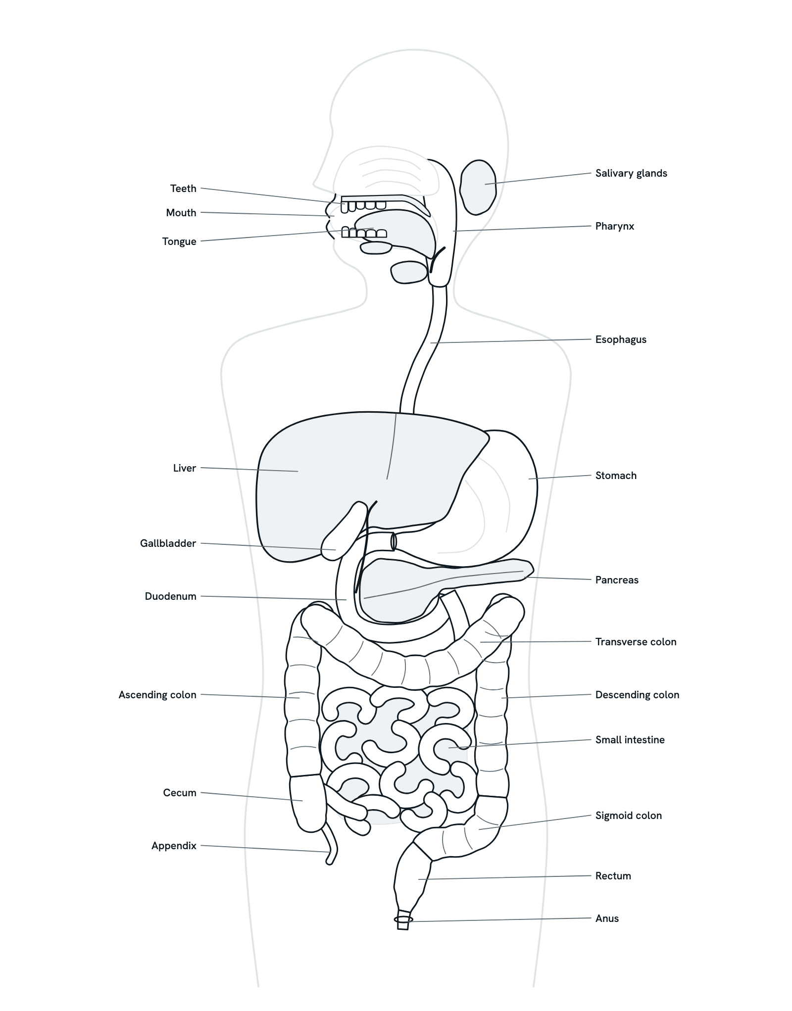
The digestive tract is one long tube from the mouth to the anus. The liver, gallbladder, pancreas and salivary glands pour digestive juices into it. Press any part to see what it does, or follow a meal through in order. Front view of the trunk, with the head turned to show the mouth and throat. Not to scale.
Also called Human digestive system or Gastrointestinal system.
Press any part of the diagram, or pick one here, to see what it is and what it does.
Download the diagramChecked against its sources on 29 October 2026.
A tour
A tour of the digestive system
01The digestive system at a glance
The digestive tract is a long, twisting tube: mouth, pharynx, esophagus, stomach, small intestine, large intestine and anus. The salivary glands, liver, gallbladder and pancreas lie outside it and send their juices in. Together they break food into parts small enough to be absorbed and used.
02Chewing and saliva
The teeth cut and grind food while the tongue mixes it with saliva from the salivary glands. Saliva moistens the food, and its amylase begins to break starch down into sugars. The chewed food becomes a soft mass, the bolus, ready to swallow.
03Swallowing
The tongue pushes the bolus back into the pharynx, and from here swallowing is automatic: the soft palate rises to seal off the nose and the epiglottis folds down over the larynx. Peristalsis carries the bolus down the esophagus, and the lower esophageal sphincter relaxes to let it into the stomach.
04The stomach
The stomach's muscular wall churns food with gastric juice. Its acid activates pepsin, an enzyme that begins the digestion of protein, while a layer of mucus protects the lining from the acid. The result is a thick liquid, chyme, which the pyloric sphincter lets into the duodenum a little at a time.
05Bile and pancreatic juice
In the duodenum, chyme meets two juices. Bile, made by the liver and stored in the gallbladder, breaks fat into tiny droplets that enzymes can reach. Pancreatic juice brings enzymes for carbohydrates, fats and proteins, and bicarbonate that neutralises the acid from the stomach.
06Absorption in the small intestine
Enzymes from the pancreas and the intestinal lining finish digestion. Folds, finger-like villi and the microvilli on their cells give the wall a very large surface. Sugars, amino acids, salts and water pass into the blood, which carries them first to the liver; most digested fat is taken up into the lymph.
07Water back in the large intestine
What is left passes from the ileum into the cecum. The large intestine makes no digestive enzymes: it absorbs water and salts, turning liquid waste into stool, while its bacteria break down some of the remaining material and make vitamin K. Peristalsis moves the stool on toward the rectum.
08Elimination
Stool is stored in the rectum until a bowel movement. Two rings of muscle keep the anus closed: the inner anal sphincter works on its own, and the outer one is under your control, so you can choose when to pass stool.
Parts
What each part of the digestive system does
- Mouth
- The start of the tract, where food is taken in, chewed and mixed with saliva. The hard and soft palate form its roof and separate it from the nasal cavity above.
- Teeth
- Cut, tear and grind food into smaller pieces, the mechanical first step of digestion, so that enzymes can work on it faster. A full adult set has 32 teeth; a child's first set has 20.
- Tongue
- A muscular organ that moves food round the mouth while it is chewed, then pushes the soft chewed mass, the bolus, back into the pharynx to be swallowed. It also carries the taste buds.
- Salivary glands
- Three pairs of large glands (parotid, submandibular and sublingual) and many small ones. Saliva moistens food so it can be swallowed, and its amylase starts the digestion of starch.
- Pharynx
- The throat, shared by air and food. When you swallow, the soft palate rises to close off the nose and the epiglottis folds down over the larynx, so food goes into the esophagus.
- Esophagus
- A muscular tube behind the trachea. Waves of muscle contraction, peristalsis, carry food down to the stomach; a ring of muscle at its lower end keeps stomach contents from flowing back. It makes mucus but no digestive enzymes.
- Stomach
- A muscular bag that churns food with gastric juice. Its acid activates pepsin, an enzyme that starts the digestion of protein, while mucus protects the lining. The pyloric sphincter lets the result, chyme, out a little at a time.
- Liver
- The largest gland in the body. For digestion it makes bile, which breaks fat into tiny droplets. It also receives the blood from the gut and processes the nutrients absorbed into it.
- Gallbladder and bile duct
- A pear-shaped sac on the underside of the liver that stores bile between meals. When you eat, it squeezes bile down the bile duct into the duodenum.
- Pancreas
- A gland behind the stomach. Its juice, carried by the pancreatic duct to the duodenum, holds enzymes for carbohydrates, fats and proteins, and bicarbonate that neutralises stomach acid. Its islets release insulin and glucagon into the blood.
- Duodenum
- The first, shortest and widest part of the small intestine, a C-shaped curve round the head of the pancreas. Chyme from the stomach meets bile and pancreatic juice here.
- Small intestine
- A long, coiled tube in three parts: the duodenum, jejunum and ileum. Digestion is completed here, and most nutrients are absorbed through its lining, whose folds, villi and microvilli give it a very large surface.
- Cecum
- The pouch at the start of the large intestine, low on the right side of the body. The ileum empties into it at the ileocecal valve, and the appendix is attached to it.
- Appendix
- A narrow, finger-shaped pouch attached to the cecum. Its wall holds lymphoid tissue that takes part in immunity, but its exact role is still debated. Inflammation of it is appendicitis.
- Colon
- The main length of the large intestine: ascending, transverse, descending and sigmoid. It absorbs water and salts, its bacteria break down some of what is left, and peristalsis moves the waste on as stool.
- Rectum
- The part of the large intestine between the sigmoid colon and the anal canal, following the curve of the sacrum. It stores stool until a bowel movement.
- Anus
- The opening at the end of the tract, kept closed by two rings of muscle: an inner sphincter that works on its own, and an outer one that is under your control.
Dictionary
The terms to know
Practise it
The quizzes and cards that fit
- Human Anatomy quizzes
- Shoulder and Rotator Cuff15 questions
- Knee Joint15 questions
- Arteries and Veins15 questions
- Physiology quizzes
- Exercise Physiology15 questions
- Puberty15 questions
- Blood Clotting15 questions
- Food Nutrition quizzes
- The Digestive System and Nutrient Absorption15 questions
- Energy Metabolism and Caloric Balance10 questions
- Protein Synthesis and Amino Acid Metabolism15 questions
- Human Anatomy flashcards
The subject's terms as a ready-made deck, free to flip through.
Next to it
More diagrams
Sources
Where the facts come from
- NIDDK: Your Digestive System and How It Works
- NCI SEER Training Modules: Regions of the Digestive System
- NCI SEER Training Modules: Accessory Organs
- StatPearls: Physiology, Digestion (NCBI Bookshelf)
- StatPearls: Anatomy, Abdomen and Pelvis, Duodenum (NCBI Bookshelf)
- StatPearls: Anatomy, Abdomen and Pelvis, Appendix (NCBI Bookshelf)
- NIDDK: Definition and Facts for Pancreatitis
Drawn and written by biofideX from these sources.