Nephron Diagram
How a nephron makes urine: filtration at the glomerulus, reabsorption along the tubule, the loop of Henle, and the hormones that set the final volume.
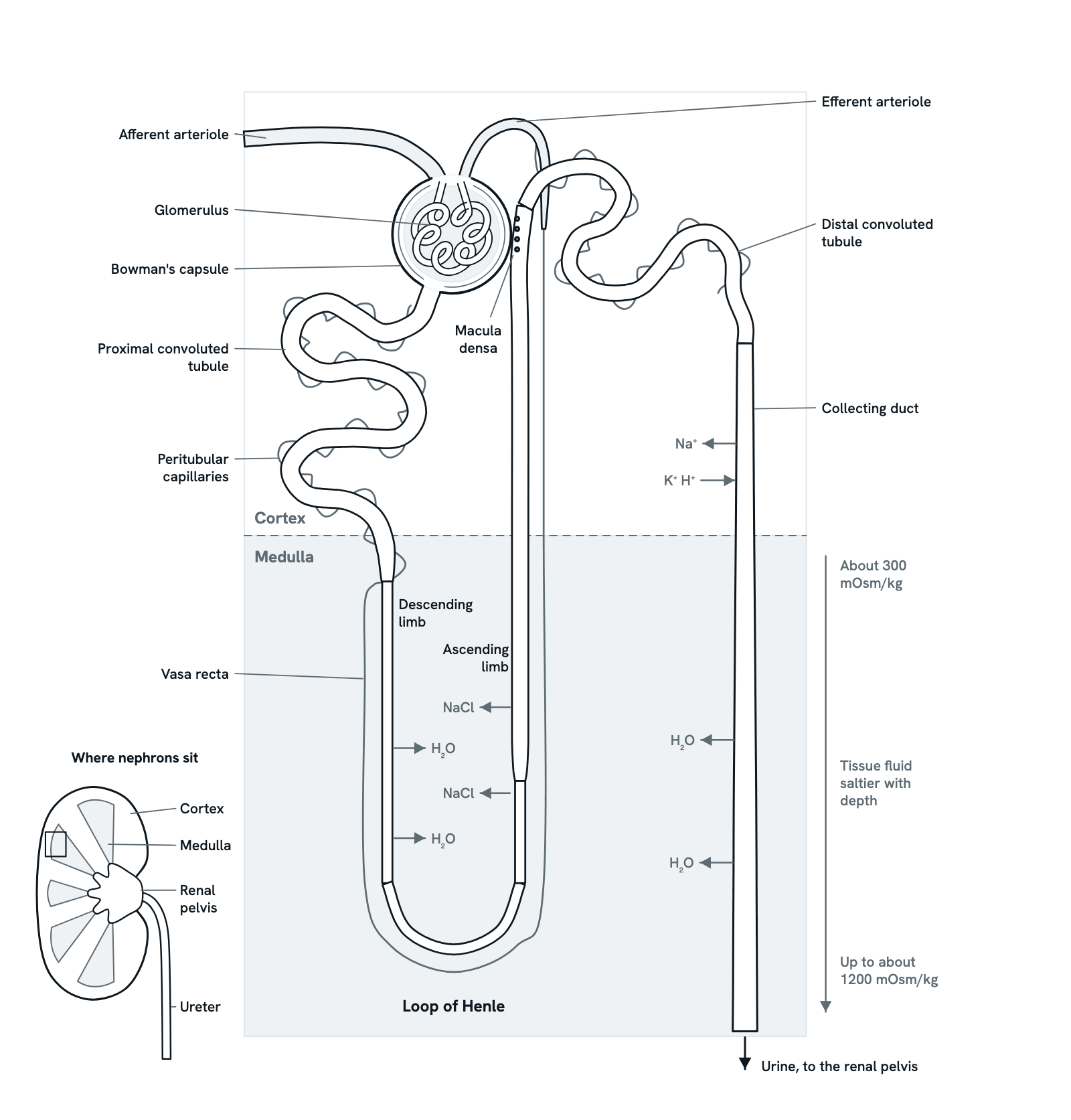
The nephron is the kidney's working unit. Blood is filtered at the glomerulus, and the tubule then takes back almost everything the body needs and adds what it must lose. Step through it in order, or press any part. A simplified nephron in section, not to scale; arrows show water and salt moving.
Also called Kidney nephron or Structure of a nephron.
Step 1 of 9
The nephron at a glance
Each kidney has around a million nephrons, though the number varies a lot between people. Each one filters blood at its glomerulus, then its tubule takes back what the body needs (reabsorption) and adds what it must lose (secretion). What is left at the end is urine.
Use ← and → to move between steps.
Step 1 of 9: The nephron at a glance
Download the diagramChecked against its sources on 29 October 2026.
Step by step
The steps of the Nephron
01The nephron at a glance
Each kidney has around a million nephrons, though the number varies a lot between people. Each one filters blood at its glomerulus, then its tubule takes back what the body needs (reabsorption) and adds what it must lose (secretion). What is left at the end is urine.
02Filtration at the glomerulus
Blood enters the glomerulus through the afferent arteriole and leaves through the efferent arteriole. The pressure in the glomerular capillaries pushes water and small solutes (salts, glucose, amino acids, urea) through their walls into Bowman's capsule, while blood cells and most proteins stay behind. Together the kidneys filter about 180 litres of fluid a day, about 120 mL a minute.
03Reabsorption in the proximal tubule
The proximal convoluted tubule takes back about two thirds of the filtered sodium and water, and nearly all the glucose and amino acids. Sodium is pumped out of the tubule cells by active transport, and water follows by osmosis. What it reclaims passes into the peritubular capillaries.
04The descending limb: water out
The descending limb runs down into the medulla, where the tissue fluid grows saltier with depth. Its wall lets water out but not salt, so water leaves by osmosis and the fluid inside becomes more concentrated as it reaches the bend.
05The ascending limb: salt out
The ascending limb lets no water through. Its thick part pumps sodium and chloride out into the medulla, which keeps the medulla salty: from about 300 mOsm/kg near the cortex to as much as about 1200 mOsm/kg deep down when the body is saving water. The fluid inside becomes more dilute as it rises.
06The macula densa checks the flow
Where the thick ascending limb touches its own glomerulus, the macula densa senses how much salt is passing. When too much arrives, the afferent arteriole narrows and filtration falls. When too little arrives, nearby cells release renin, which leads to angiotensin II and aldosterone.
07The distal tubule and aldosterone
The distal convoluted tubule reabsorbs more sodium and chloride, and calcium under parathyroid hormone. In its last part and in the collecting duct, aldosterone makes cells take back more sodium and secrete potassium, while other cells secrete hydrogen ions, which helps keep the blood's pH steady.
08The collecting duct and ADH
The collecting duct carries fluid from several nephrons down through the salty medulla. Antidiuretic hormone (ADH) puts water channels, aquaporin-2, into its cells, so water leaves and the urine becomes concentrated. With little ADH, the duct stays closed to water and a large volume of dilute urine passes.
09Urine leaves the kidney
At the tips of the pyramids the collecting ducts empty into the calyces, which drain into the renal pelvis and the ureter, and so to the bladder. Of the 180 litres filtered each day, only about 1 to 2 litres leave as urine.
Parts
What each part of the nephron does
- Afferent arteriole
- The small artery that brings blood into the glomerulus. By narrowing or widening, it sets how much blood, and so how much pressure, reaches the glomerular capillaries.
- Glomerulus
- A tuft of capillary loops inside Bowman's capsule. Their walls let water and small dissolved substances through into the capsule but hold back blood cells and most proteins.
- Bowman's capsule
- A double-layered cup of epithelium round the glomerulus. Its inner layer of podocytes wraps the capillaries; the space between the layers collects the filtrate and leads into the proximal tubule.
- Efferent arteriole
- Carries blood away from the glomerulus, then divides again into the peritubular capillaries and the vasa recta, which take back what the tubule reabsorbs. Angiotensin II narrows it, which helps keep filtration up when blood flow falls.
- Proximal convoluted tubule
- The first coiled part of the tubule, in the cortex. It takes back about two thirds of the filtered sodium and water, and nearly all the glucose and amino acids.
- Descending limb of the loop of Henle
- Runs down into the medulla. It lets water out but holds on to salt, so the fluid inside grows more concentrated as it goes deeper into the salty medulla.
- Loop of Henle
- The hairpin that dips from the cortex into the medulla and back. Its two limbs together build and keep up the salty medulla that lets the kidney make concentrated urine.
- Ascending limb of the loop of Henle
- Runs back up to the cortex and lets no water out. Its thick part pumps sodium and chloride out into the medulla, so the fluid inside becomes more dilute as it rises.
- Macula densa
- A patch of tightly packed cells at the end of the thick ascending limb, where it touches the vascular pole of its own glomerulus. With the renin-making cells of the afferent arteriole and the mesangial cells beside them, it forms the juxtaglomerular apparatus, which senses the salt in the tubule and adjusts filtration.
- Distal convoluted tubule
- A short coiled tubule back in the cortex. It reabsorbs more sodium and chloride, and calcium under the control of parathyroid hormone. In its last part, aldosterone begins to act.
- Collecting duct
- Takes fluid from several nephrons down through the medulla. Aldosterone makes it reabsorb sodium and secrete potassium; antidiuretic hormone opens water channels so water leaves for the salty medulla.
- Peritubular capillaries
- The second capillary bed, fed by the efferent arteriole. They wind round the tubules in the cortex and take back what the tubule reabsorbs. In the medulla, long loops called the vasa recta run beside the loop of Henle.
- Renal cortex
- The outer layer of the kidney. It holds the glomeruli, Bowman's capsules and the convoluted tubules of every nephron.
- Renal medulla
- The inner layer, arranged in cone-shaped pyramids. Loops of Henle and collecting ducts run through it, and its tissue fluid gets saltier with depth, from about 300 to as much as about 1200 mOsm/kg.
- Renal pelvis and ureter
- Urine drains from the tips of the pyramids into cups called calyces, then into the funnel-shaped renal pelvis, and down the ureter to the bladder.
Dictionary
The terms to know
Practise it
The quizzes and cards that fit
- Physiology quizzes
- Exercise Physiology15 questions
- Puberty15 questions
- Blood Clotting15 questions
- Human Anatomy quizzes
- Shoulder and Rotator Cuff15 questions
- Knee Joint15 questions
- Arteries and Veins15 questions
- Medicine quizzes
- Labor and Delivery15 questions
- IV Fluids15 questions
- Sepsis15 questions
- Physiology flashcards
The subject's terms as a ready-made deck, free to flip through.
- Normal Lab Values
Typical adult ranges for blood, urine and spinal fluid tests, in conventional and SI units, with a source for every value.
- mg/dL to mmol/L Converter
Convert glucose, cholesterol, creatinine, HbA1c and other results between conventional and SI units.
Next to it
More diagrams
Sources
Where the facts come from
- StatPearls: Physiology, Renal (NCBI Bookshelf)
- StatPearls: Physiology, Glomerular Filtration Rate (NCBI Bookshelf)
- StatPearls: Physiology, Renal Blood Flow and Filtration (NCBI Bookshelf)
- StatPearls: Anatomy, Abdomen and Pelvis: Kidneys (NCBI Bookshelf)
- StatPearls: Anatomy, Abdomen and Pelvis: Kidney Collecting Ducts (NCBI Bookshelf)
- StatPearls: Physiology, Aldosterone (NCBI Bookshelf)
- StatPearls: Physiology, Vasopressin (NCBI Bookshelf)
- Sands and Layton, The Physiology of Urinary Concentration: an Update, Seminars in Nephrology, 2009 (PubMed Central)
- Peti-Peterdi and Harris, Macula Densa Sensing and Signaling Mechanisms of Renin Release, Journal of the American Society of Nephrology, 2010 (PubMed Central)
- NIDDK: Your Kidneys and How They Work
- NCI SEER Training: Kidneys
Drawn and written by biofideX from these sources.