Coagulation Cascade Diagram
How the coagulation cascade turns fibrinogen into fibrin: the intrinsic, extrinsic and common pathways, the PT and aPTT, and the drugs that act on it.
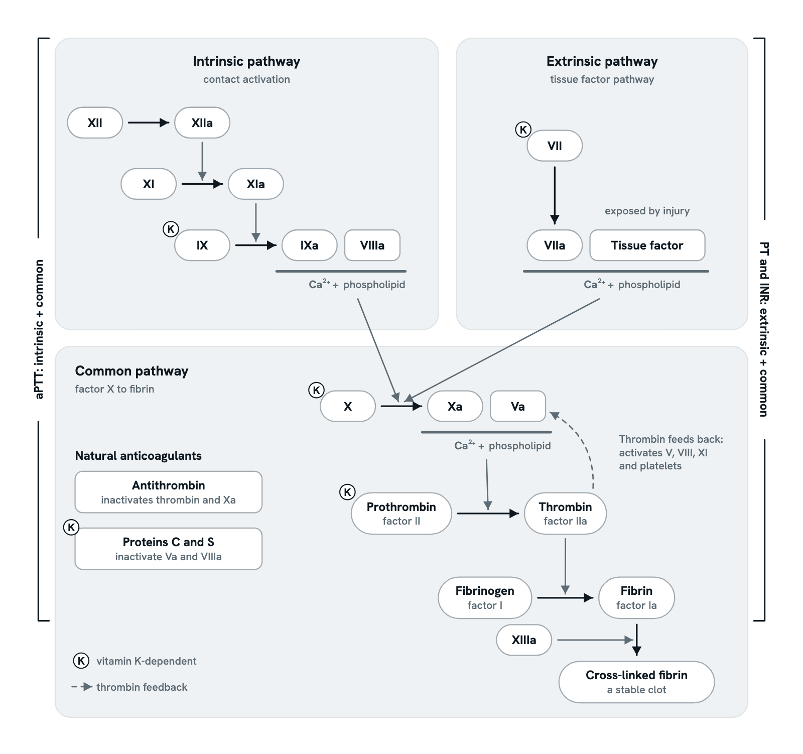
Clotting factors circulate as inactive precursors, and each one, once activated, activates the next, until thrombin turns fibrinogen into a fibrin mesh. This is the classic three-pathway picture, bracketed by the lab test that reads each arm. Step through it, from the pathways to the cell-based model, warfarin, heparin and hemophilia, or press any factor.
Also called Clotting cascade, Coagulation pathway or Blood clotting cascade.
Step 1 of 10
Three pathways, one clot
Clotting factors circulate as inactive precursors. Once activated, each one activates the next, marked by an "a" after its numeral. In the classic picture, the intrinsic and extrinsic pathways both activate factor X, and the common pathway then makes thrombin, which turns fibrinogen into fibrin.
Use ← and → to move between steps.
Step 1 of 10: Three pathways, one clot
Download the diagramChecked against its sources on 29 October 2026.
Step by step
The steps of the Coagulation Cascade
01Three pathways, one clot
Clotting factors circulate as inactive precursors. Once activated, each one activates the next, marked by an "a" after its numeral. In the classic picture, the intrinsic and extrinsic pathways both activate factor X, and the common pathway then makes thrombin, which turns fibrinogen into fibrin.
02The intrinsic pathway
In the test tube, factor XII is activated by contact with a negatively charged surface. XIIa activates XI, and XIa activates IX. Factor IXa joins its cofactor VIIIa on a phospholipid surface with calcium, and this complex, the intrinsic tenase, activates factor X.
03The extrinsic pathway
Tissue factor sits in the membranes of cells beneath the lining of blood vessels. When a vessel is damaged, blood reaches it, and factor VII binds it and becomes VIIa. Tissue factor with VIIa and calcium activates factor X. With fewer steps, it is the shorter arm.
04The common pathway makes thrombin
Both arms meet at factor X. Factor Xa joins its cofactor Va on a phospholipid surface with calcium, in a complex called prothrombinase. Prothrombinase cuts prothrombin (factor II) to release thrombin (factor IIa).
05Thrombin turns fibrinogen into fibrin
Thrombin cuts soluble fibrinogen into fibrin, which joins into strands that bind the platelet plug together. Thrombin also activates factor XIII, and XIIIa cross-links the strands into a firm, stable clot.
06What the PT and aPTT read
The PT adds tissue factor to plasma and reads the extrinsic and common pathways; the INR corrects it for the reagent, so results compare between labs. The aPTT adds a contact activator and reads the intrinsic and common pathways. Both stop the clock when fibrin forms, so neither picks up a lack of factor XIII.
07In the body: the cell-based model
The split into two pathways describes the test tube better than the body. In the body, tissue factor with VIIa starts clotting, activating X and IX and making a little thrombin. That thrombin activates platelets and factors V, VIII and XI, and the complexes on the platelet surface then make thrombin in a burst. People who lack factor XII do not bleed abnormally.
08Vitamin K and warfarin
The liver needs vitamin K to finish factors II, VII, IX and X, and the anticoagulant proteins C and S. Warfarin blocks the recycling of vitamin K, so the liver releases fewer working factors. Warfarin is monitored with the PT and INR; for most patients the target INR is 2 to 3.
09Heparin works through antithrombin
Antithrombin is a natural inhibitor of thrombin and factor Xa, and of IXa, XIa and XIIa. Heparin binds antithrombin and activates it. Unfractionated heparin is monitored with the aPTT; low-molecular-weight heparin does not need routine aPTT checks.
10Hemophilia A and B
Hemophilia A is a lack of factor VIII, and hemophilia B a lack of factor IX. Both are X-linked recessive, so they mostly affect males, and both lengthen the aPTT while the PT stays normal. The extrinsic arm cannot make up for them: it starts clotting, but the burst of thrombin on platelets needs IXa with VIIIa.
Parts
What each part of the coagulation cascade does
- Factor XII (Hageman factor)
- Starts the intrinsic pathway in the test tube, when it touches a negatively charged surface such as the kaolin of the aPTT. People who lack it do not bleed abnormally, a sign that it matters little for clotting in the body.
- Factor XI
- Activated by XIIa in the classic picture, and in the body by thrombin too. Factor XIa activates factor IX. People who lack factor XI (hemophilia C) usually bleed only mildly.
- Factor IX (Christmas factor)
- A vitamin K-dependent enzyme, activated by XIa and, in the body, by tissue factor with VIIa. With its cofactor VIIIa, factor IXa activates factor X. A lack of factor IX causes hemophilia B.
- Factor VIII
- A cofactor rather than an enzyme. Activated by thrombin, VIIIa binds factor IXa on a phospholipid surface with calcium and makes it activate factor X far faster. A lack of factor VIII causes hemophilia A.
- Tissue factor (factor III)
- A membrane protein of cells beneath the lining of blood vessels, out of the blood's reach until a vessel is damaged. Bound to factor VIIa, with calcium, it activates factor X, and factor IX too. It starts clotting in the body.
- Factor VII (stable factor)
- A vitamin K-dependent enzyme that becomes VIIa on tissue factor. It has the shortest half-life of the clotting factors, so it falls first when the liver makes fewer of them, and the PT is the first test to lengthen.
- Factor X (Stuart-Prower factor)
- Where the two arms meet: IXa with VIIIa, or tissue factor with VIIa, activates it. Factor Xa joins its cofactor Va to form prothrombinase, which turns prothrombin into thrombin. It is vitamin K-dependent.
- Factor V
- The cofactor of factor Xa. Activated by thrombin, Va binds Xa on a phospholipid surface with calcium, forming prothrombinase. Activated protein C, with protein S, switches it off.
- Prothrombin (factor II)
- A vitamin K-dependent protein made in the liver: the inactive precursor of thrombin. Prothrombinase cuts it to release thrombin.
- Thrombin (factor IIa)
- The cascade's main product: the enzyme that turns fibrinogen into fibrin and activates factor XIII. It also feeds back on the cascade, so a little thrombin leads to much more.
- Thrombin feedback
- Thrombin activates factors V, VIII and XI and platelets, so the cofactors and the platelet surface for the complexes are ready. The dashed arrow onto Va stands for all of these.
- Fibrinogen (factor I)
- A large, soluble plasma protein made by the liver. Thrombin cuts it into fibrin units that join into strands.
- Fibrin
- The strands formed when thrombin cuts fibrinogen. They bind platelets together into a firmer plug. The PT and aPTT stop the clock when fibrin first forms in the tube.
- Factor XIII (fibrin-stabilising factor)
- Activated by thrombin, XIIIa joins fibrin strands to one another with covalent bonds. Neither the PT nor the aPTT measures it, because both stop when fibrin first forms.
- Cross-linked fibrin
- The finished mesh of cross-linked fibrin that holds the platelet plug in place: the end of secondary hemostasis.
- Calcium (factor IV)
- Calcium ions hold the vitamin K-dependent factors to the phospholipid surface, so the complexes can form. Tubes for clotting tests contain citrate, which binds calcium; the lab adds calcium back to start the test.
- Phospholipid surface
- The membrane the complexes assemble on, in the body mainly that of activated platelets, which expose the negatively charged phospholipid phosphatidylserine. In the aPTT, a phospholipid reagent stands in for it.
- Vitamin K-dependent factors
- The liver needs vitamin K to finish factors II, VII, IX and X and proteins C and S, adding the groups that bind calcium. Warfarin blocks the recycling of vitamin K, so fewer working factors are made.
- Antithrombin
- A natural inhibitor in the plasma that inactivates thrombin and factor Xa, and also IXa, XIa and XIIa. Heparin binds antithrombin and makes it far more active.
- Proteins C and S
- Natural anticoagulants, both vitamin K-dependent. Thrombin bound to thrombomodulin on the vessel lining activates protein C, which, with protein S as its cofactor, inactivates factors Va and VIIIa.
- PT and INR (prothrombin time)
- Plasma is timed until it clots after tissue factor, phospholipid and calcium are added: about 11 to 15 seconds, though each lab sets its own range. It reads factors VII, X, V and II and fibrinogen. The INR corrects for the reagent, so results compare between labs.
- aPTT (activated partial thromboplastin time)
- Plasma is timed until it clots after a contact activator, phospholipid and calcium are added: about 25 to 40 seconds, though each lab sets its own range. It reads every factor except VII and XIII, and monitors unfractionated heparin.
Dictionary
The terms to know
Practise it
The quizzes and cards that fit
- Physiology quizzes
- Exercise Physiology15 questions
- Puberty15 questions
- Blood Clotting15 questions
- Medicine quizzes
- Labor and Delivery15 questions
- IV Fluids15 questions
- Sepsis15 questions
- Pathology quizzes
- Hematopathology: Anemias & WBC Disorders15 questions
- Cell Injury, Death & Adaptations10 questions
- Cardiovascular Pathology15 questions
- Physiology flashcards
The subject's terms as a ready-made deck, free to flip through.
- Normal Lab Values
Typical adult ranges for blood, urine and spinal fluid tests, in conventional and SI units, with a source for every value.
Next to it
More diagrams
Sources
Where the facts come from
- StatPearls: Physiology, Coagulation Pathways (NCBI Bookshelf)
- StatPearls: Biochemistry, Clotting Factors (NCBI Bookshelf)
- StatPearls: Prothrombin Time (NCBI Bookshelf)
- StatPearls: Partial Thromboplastin Time (NCBI Bookshelf)
- Palta, Saroa and Palta: Overview of the coagulation system, Indian Journal of Anaesthesia 2014 (PMC)
Drawn and written by biofideX from these sources.