DNA Structure Diagram
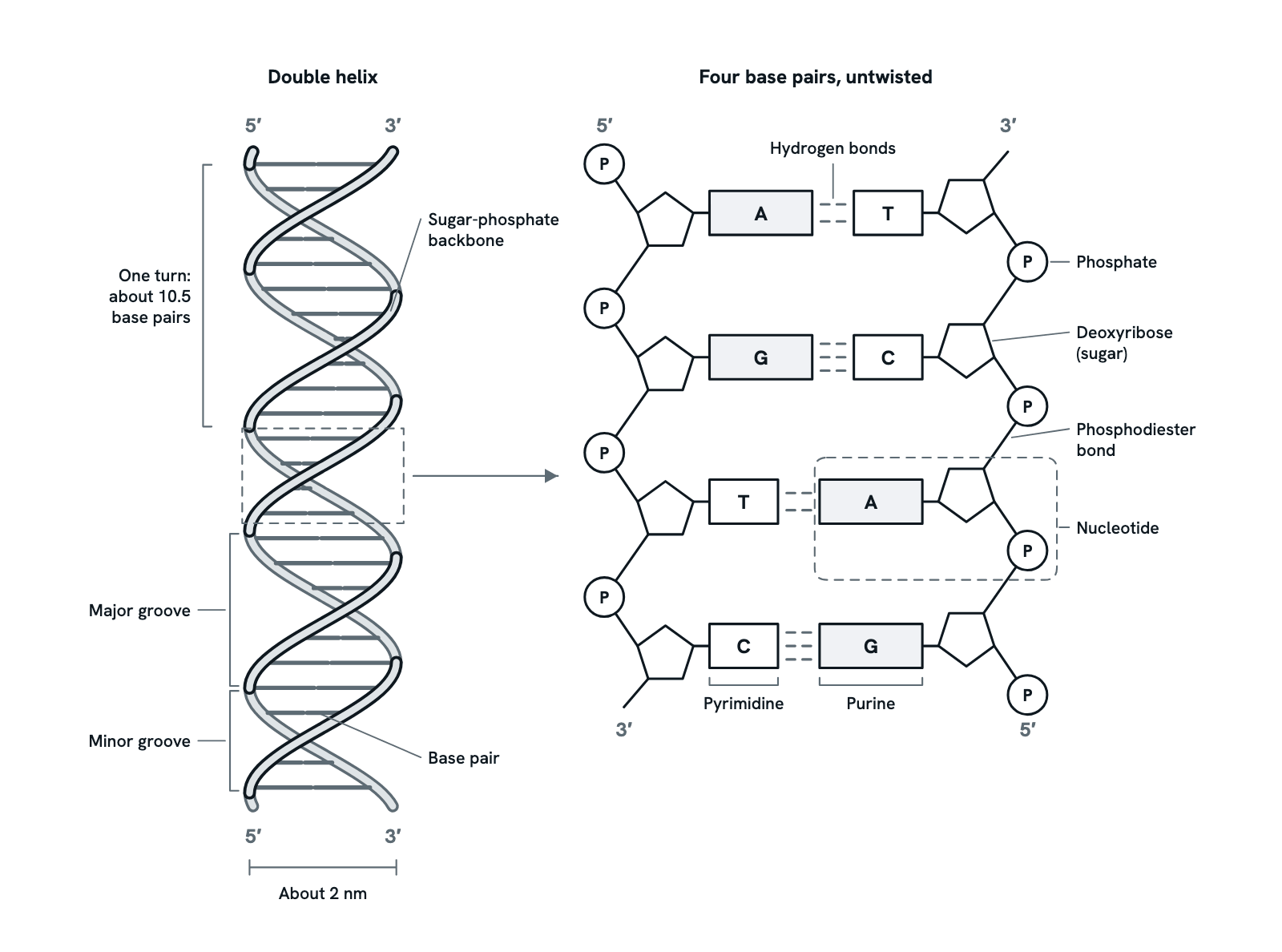
The structure of DNA: nucleotides, the sugar-phosphate backbone, base pairs and hydrogen bonds, antiparallel strands and the grooves.
DNA is two strands of nucleotides wound round each other in a double helix. On the left is the helix seen from the side; on the right, four of its base pairs taken out, untwisted and enlarged, so each part of a nucleotide can be seen. Press any part to see what it is, or take the tour in order. Not to scale.
Also called DNA double helix or Structure of DNA.
Press any part of the diagram, or pick one here, to see what it is and what it does.
Download the diagramChecked against its sources on 29 October 2026.
A tour
A tour of the dna structure
01DNA structure at a glance
DNA is made of two long strands of nucleotides. The strands are held together by pairs of bases in the middle and wound round each other in a double helix, like a twisted ladder: the sugar-phosphate backbones are the sides and the base pairs the rungs.
02The nucleotide
Each strand is a chain of nucleotides. A nucleotide has three parts: a phosphate group, a five-carbon sugar called deoxyribose, and a nitrogen-containing base, which is adenine (A), thymine (T), guanine (G) or cytosine (C). Only the base differs from one nucleotide to the next.
03The sugar-phosphate backbone
The nucleotides of a strand are joined by phosphodiester bonds: a phosphate links the 3′ carbon of one sugar to the 5′ carbon of the next. Sugar and phosphate alternate along the strand, forming a strong backbone on the outside of the helix, with the bases pointing inwards.
04The bases: purines and pyrimidines
Adenine and guanine are purines, with two rings. Thymine and cytosine are pyrimidines, with one ring. In every base pair a purine faces a pyrimidine, so each pair is about the same width and the two backbones stay an even distance apart.
05Complementary base pairing
A always pairs with T, held by two hydrogen bonds, and G with C, held by three. So the sequence of one strand fixes the sequence of the other, and a DNA molecule has as much A as T and as much G as C. Each strand can serve as a template for copying the other.
06Antiparallel strands
Each strand has a direction, from its 5′ end to its 3′ end. The two strands of the helix run in opposite directions: where one reads 5′ to 3′ downwards, its partner reads 5′ to 3′ upwards. In the enlarged view, the sugars of the two strands point opposite ways.
07The double helix
The two backbones wind round each other in a right-handed helix, with the base pairs stacked flat inside it. In B-DNA, the usual form in cells, the helix is about 2 nm wide, each base pair sits 0.34 nm above the last, and it makes a full turn about every 10.5 base pairs.
08Major and minor grooves
The two backbones are not evenly spaced round the helix, so two grooves of different widths spiral along it: the major groove and the minor groove. The edges of the bases are exposed in the grooves, and proteins such as transcription factors read the sequence there, mostly in the major groove, without opening the helix.
Parts
What each part of the dna structure does
- Sugar-phosphate backbone
- The outside of each strand: sugars and phosphates in turn, joined by strong covalent bonds. The bases point inwards from it, into the middle of the helix.
- Base pair
- Two bases, one from each strand, held together by hydrogen bonds: A with T, G with C. Each pair is a rung of the twisted ladder, and every pair is about the same width.
- Nucleotide
- The unit DNA is built from: a phosphate group, a deoxyribose sugar and one of four bases (A, T, G or C). A strand is a chain of nucleotides.
- Phosphate group
- Joined to the 5′ carbon of its own sugar and, in the chain, to the 3′ carbon of the sugar before it, so that sugar and phosphate alternate along the strand.
- Deoxyribose
- The five-carbon sugar of DNA, drawn as a pentagon (four carbons and an oxygen make the ring). It lacks the hydroxyl group on its 2′ carbon that ribose in RNA has. The base is joined to its 1′ carbon.
- Phosphodiester bond
- The link between nucleotides in a strand: a phosphate joins the 3′ carbon of one sugar to the 5′ carbon of the next. These covalent bonds make the backbone strong.
- Purines (A and G)
- Adenine and guanine are purines, larger bases with two rings, drawn here as the long grey boxes. Each pairs with a pyrimidine on the other strand.
- Pyrimidines (T and C)
- Thymine and cytosine are pyrimidines, smaller bases with one ring, drawn as the short white boxes. A purine and a pyrimidine together always span the same width.
- Hydrogen bonds
- Weak bonds between the bases of a pair: two between A and T, three between G and C. One by one they are weak, so the strands can be parted for copying and reading.
- 5′ and 3′ ends
- Each strand has a 5′ end, with a phosphate on the 5′ carbon of its last sugar, and a 3′ end, with a free hydroxyl on the 3′ carbon. The two strands run in opposite directions.
- One turn of the helix
- In B-DNA, the usual form in cells, each base pair sits 0.34 nm above the last and the helix turns once about every 10.5 base pairs (about 10 in the DNA fibres studied by X-ray).
- Width of the helix
- The double helix is about 2 nm across. Because a two-ring purine always pairs with a one-ring pyrimidine, the width stays about the same all along the molecule.
- Major groove
- The wider of the two grooves that spiral round the helix. The edges of the bases are exposed in it, and most proteins that read a DNA sequence bind here.
- Minor groove
- The narrower groove, where the two backbones run closest together. The edges of the bases are exposed here too, but they are harder to tell apart than in the major groove.
Dictionary
The terms to know
Practise it
The quizzes and cards that fit
- Molecular Biology quizzes
- Epigenetics & Chromatin Remodeling20 questions
- DNA Replication Machinery15 questions
- Recombinant DNA Technology15 questions
- Genetics quizzes
- Incomplete Dominance and Codominance15 questions
- Pedigree Analysis15 questions
- Blood Types15 questions
- Biochemistry quizzes
- Photosynthesis and Cellular Respiration15 questions
- Molarity and Dilutions15 questions
- ATP and Cellular Energy15 questions
- Molecular Biology flashcards
The subject's terms as a ready-made deck, free to flip through.
- Reverse Complement
The reverse complement, complement or reverse of a DNA or RNA sequence, or DNA written as RNA, from bare letters or FASTA.
- GC Content Calculator
GC content, base counts, length and molecular weight of a DNA or RNA sequence, with GC along the sequence in a sliding window.
Next to it
More diagrams
Sources
Where the facts come from
- Alberts and others, Molecular Biology of the Cell, 4th edition: The Structure and Function of DNA (NCBI Bookshelf)
- Alberts and others, Molecular Biology of the Cell, 4th edition: DNA-Binding Motifs in Gene Regulatory Proteins (NCBI Bookshelf)
- StatPearls: Biochemistry, DNA Structure (NCBI Bookshelf)
- NHGRI Talking Glossary: Double Helix
- NHGRI Talking Glossary: Base Pair
- Levitt, How many base-pairs per turn does DNA have in solution and in chromatin?, PNAS 1978 (PMC)
Drawn and written by biofideX from these sources.