Hemoglobin 3D Structure
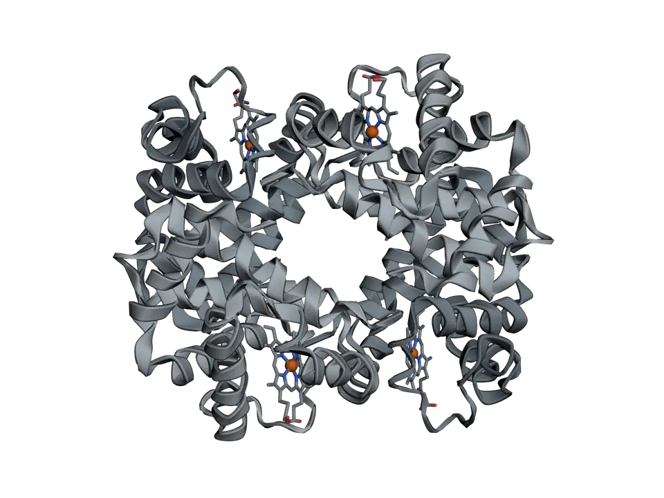
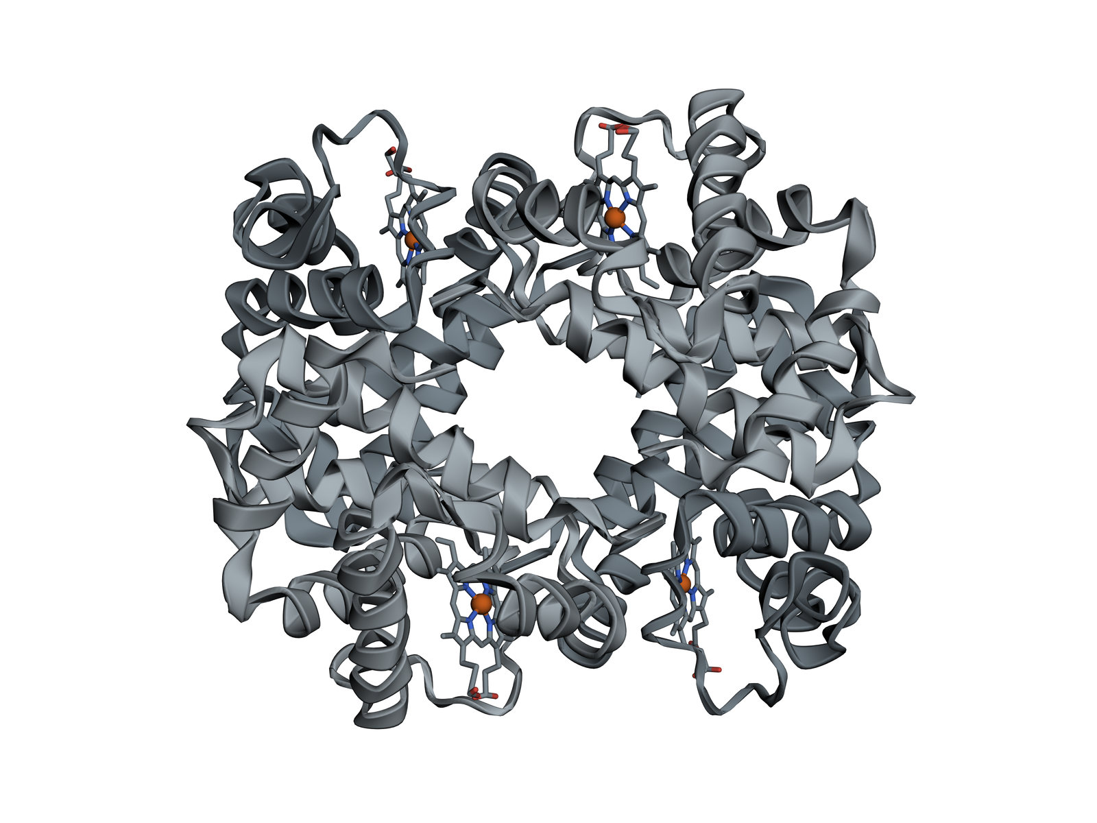
The protein that carries oxygen in red blood cells: four chains, each holding a haem group whose iron binds one oxygen molecule.
Adult haemoglobin is built from two alpha and two beta chains, each folded around a haem group with an iron at its centre. It loads oxygen in the lungs and lets it go in the tissues, and it does both well because the four chains work together. This structure, from Max Perutz's laboratory, is human haemoglobin without oxygen bound.
Also called Haemoglobin, Hb or Haemoglobin A.
View 1 of 7
Four chains, one molecule
Haemoglobin is a tetramer: four protein chains packed into a rounded molecule about 6 nanometres across. Each chain holds one haem, so one haemoglobin can carry up to four oxygen molecules.
Use ← and → to move between views. Drag the molecule to turn it; on a touch screen, press Turn it first.
View 1 of 7: Four chains, one molecule
Download the pictureDownload the atoms (PDB)Checked against its sources on 25 October 2026.
View by view
The Hemoglobin structure, explained
01Four chains, one molecule
Haemoglobin is a tetramer: four protein chains packed into a rounded molecule about 6 nanometres across. Each chain holds one haem, so one haemoglobin can carry up to four oxygen molecules.
02Two alpha chains
The two alpha chains are identical, each 141 amino acids long. Each folds into alpha helices labelled A to H, missing only a short D helix that the beta chains have: the globin fold, which myoglobin shares.
03Two beta chains
The two beta chains are 146 amino acids long. In the molecule each pairs with an alpha chain, so haemoglobin is really two alpha-beta pairs, α1β1 and α2β2, set against each other.
04A haem in every chain
Each chain wraps a haem group in a pocket lined with water-repelling amino acids. The pocket keeps water away from the iron, which keeps it in its ferrous form, able to bind oxygen, rather than oxidised to the ferric form of methaemoglobin, which cannot.
05Where oxygen binds
Close up on one haem of α1. The iron is held from below by the proximal histidine, and oxygen binds on the other side, beside the distal histidine. In this structure, without oxygen, the iron sits slightly out of the plane of the ring.
06Four chains that work together
When oxygen binds, the iron moves into the plane of the ring and pulls the proximal histidine with it. The shift spreads to the contacts between the two alpha-beta pairs, and the molecule turns from the T state, shown here, to the R state, in which the other chains bind oxygen more easily. This is why haemoglobin's oxygen binding curve is S-shaped.
07One amino acid, one disease
In sickle cell disease the sixth amino acid of each beta chain, glutamate, is replaced by valine. The change sits on the surface, and when haemoglobin S loses its oxygen it sticks to its neighbours and forms long fibres that bend red cells out of shape.
Parts
What each part does
- Alpha chain (α1)
- One of the two alpha globins, 141 amino acids folded into a bundle of alpha helices that cradles one haem.
- Beta chain (β1)
- One of the two beta globins, 146 amino acids. It folds the same way as an alpha chain and pairs tightly with α1.
- Alpha chain (α2)
- The second alpha globin, identical in sequence to α1.
- Beta chain (β2)
- The second beta globin. The β2 chain pairs with α2, and where the α1β1 pair meets the α2β2 pair, the molecule shifts as it takes up oxygen.
- Haem groups
- Four flat rings of protoporphyrin IX, one in a pocket of each chain, each holding one iron. Haem is what makes blood red.
- Iron
- The ferrous iron (Fe²⁺) at the centre of each haem, held by four nitrogens of the ring. Oxygen binds to it, one molecule per iron.
- Proximal histidine
- A histidine on each chain (α87 and β92) that holds the iron from below. When oxygen binds, the iron moves into the ring and pulls this histidine, and its helix, with it.
- Distal histidine
- A histidine on the oxygen side of each haem (α58 and β63). It does not touch the iron but forms a hydrogen bond to oxygen once bound, which helps hold it.
- Glutamate 6 of each beta chain
- The amino acid changed in sickle cell disease. Valine in its place leaves a sticky patch on the surface, and deoxygenated haemoglobin S joins into long fibres.
The structure
Where these atoms come from
- Structure
- PDB 4HHB
- What it is
- Human deoxyhaemoglobin
- Organism
- Homo sapiens
- Method
- X-ray diffraction, 1.74 Å resolution
- Deposited
- 1984
- Atoms shown
- 4,556, without water or hydrogens
Coordinates from the Protein Data Bank, free of all copyright (CC0). Water and hydrogens are left out, and the molecule is turned to face you; every atom is where the experiment placed it.
Dictionary
The terms to know
Practise it
The quizzes and cards that fit
- Physiology quizzes
- Exercise Physiology15 questions
- Puberty15 questions
- Blood Clotting15 questions
- Biochemistry quizzes
- Photosynthesis and Cellular Respiration15 questions
- Molarity and Dilutions15 questions
- ATP and Cellular Energy15 questions
- Physiology flashcards
The subject's terms as a ready-made deck, free to flip through.
Next to it
More molecules
Sources
Where the facts come from
- RCSB Protein Data Bank: 4HHB, the crystal structure of human deoxyhaemoglobin
- Fermi and others, The crystal structure of human deoxyhaemoglobin at 1.74 Å resolution, J Mol Biol 1984
- PDB-101 Molecule of the Month: Hemoglobin
- StatPearls: Physiology, Oxygen Transport And Carbon Dioxide Dissociation Curve (NCBI Bookshelf)
Written by biofideX from these sources. Drawn with 3Dmol.js (Rego and Koes, Bioinformatics 2015).