Antibody 3D Structure
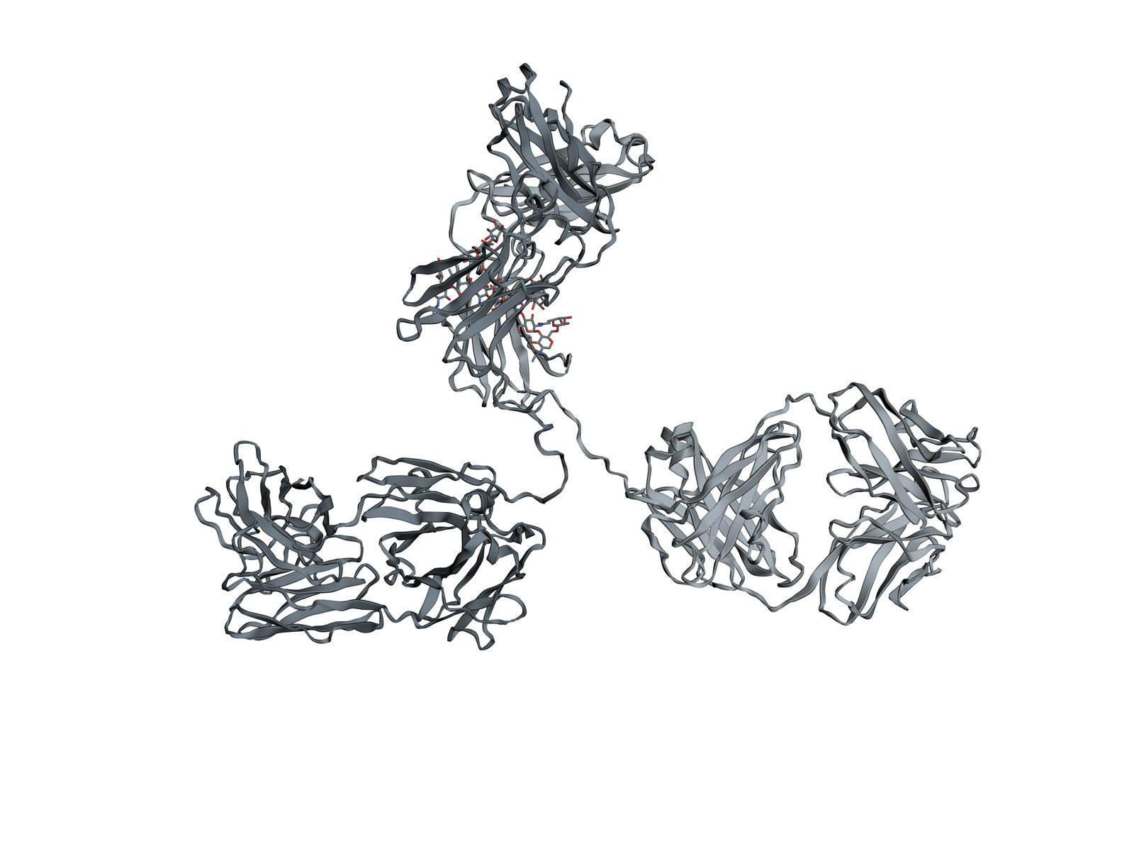
A Y-shaped protein of the immune system: two arms that bind a target, and a stem that calls in the cells and proteins that destroy it.
Antibodies are made by B cells and plasma cells. An IgG like this one is built from two identical heavy chains and two identical light chains, folded into twelve similar domains and joined by disulfide bonds. The tips of the two arms bind one shape, the antigen, and the stem tells the rest of the immune system what to do with it. This is a mouse IgG2a, one of the first whole antibodies seen in a crystal.
Also called Immunoglobulin G or IgG.
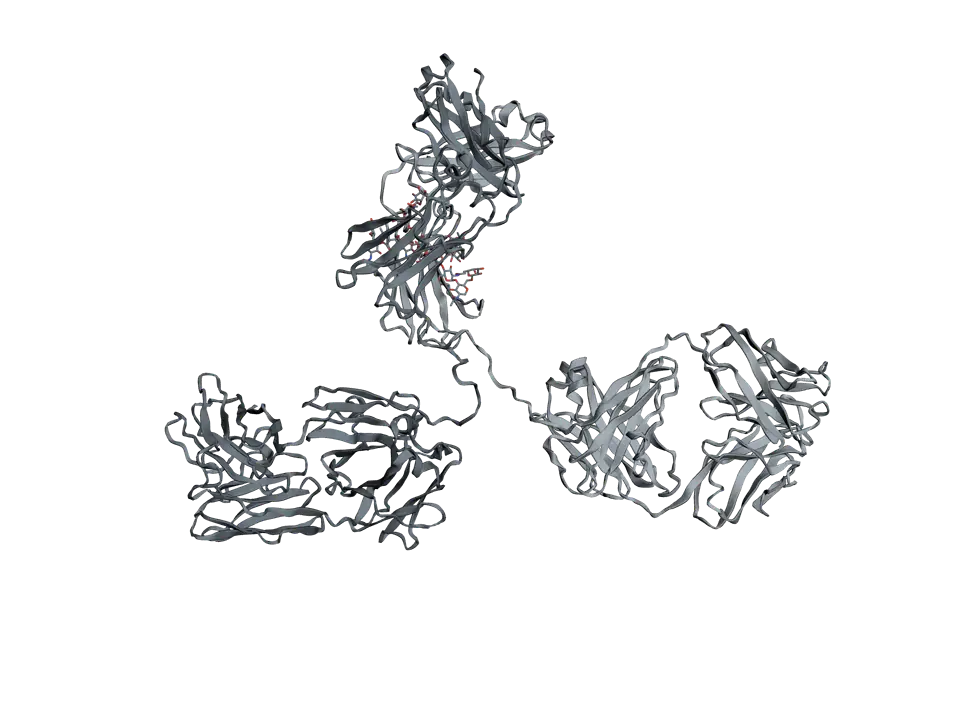
View 1 of 7
A Y-shaped protein
An IgG antibody is about 150 kilodaltons: two arms and a stem, each about the same size, joined at a hinge. In this crystal the molecule is caught in one of the many shapes its flexible hinge allows.
Use ← and → to move between views. Drag the molecule to turn it; on a touch screen, press Turn it first.
View 1 of 7: A Y-shaped protein
Download the pictureDownload the atoms (PDB)Checked against its sources on 25 October 2026.
View by view
The Antibody structure, explained
01A Y-shaped protein
An IgG antibody is about 150 kilodaltons: two arms and a stem, each about the same size, joined at a hinge. In this crystal the molecule is caught in one of the many shapes its flexible hinge allows.
02Two light chains
Each light chain, about 25 kilodaltons, folds into two domains of the same kind, the immunoglobulin fold: a sandwich of two beta sheets. In mammals a light chain is either kappa or lambda; both kinds work the same way.
03Two heavy chains
Each heavy chain, about 50 kilodaltons, folds into four domains. The heavy chain decides the class of the antibody, IgG, IgA, IgM, IgD or IgE, and with it where the antibody goes and what it does.
04Two arms that bind
Each arm, a Fab, is a whole light chain paired with the first two domains of a heavy chain. Both arms of one antibody are identical and bind the same antigen.
05The loops at the tips
At the tip of each arm, six loops form the antigen-binding site. B cells shuffle gene segments to build these loops, and mutate them further during an immune response, so the body can make antibodies against almost any shape.
06A hinge between arms and stem
Between the arms and the stem, each heavy chain runs through a short hinge held to the other by disulfide bonds. The hinge lets the arms swing apart or together, so both can grip copies of an antigen spaced differently on a surface.
07A stem that calls for help
The stem, the Fc, does not bind antigen. Once the arms have bound, the Fc is what phagocytes grab through their Fc receptors and what the first complement protein, C1q, binds, so the target is swallowed or destroyed.
Parts
What each part does
- Light chains
- Two identical chains of about 214 amino acids, each folded into two domains: one variable, at the tip of an arm, and one constant.
- Heavy chains
- Two identical chains of about 450 amino acids, each folded into four domains. They run from the tip of each arm down through the hinge into the stem.
- Fab arm
- One arm: a light chain beside the first half of a heavy chain. Fab means fragment, antigen binding, the piece the enzyme papain cuts off.
- Second Fab arm
- The other arm, the same as the first. With two arms, one antibody can hold two copies of its antigen, which helps it clump viruses and bacteria together.
- Antigen-binding loops
- Six loops at the tip of each arm, three from the light chain and three from the heavy chain, called complementarity-determining regions. Their sequence differs from one antibody to the next, and so does the shape they bind.
- Hinge
- A short, flexible stretch of each heavy chain between the arms and the stem, rich in proline. It lets the arms swing to reach targets at different distances apart.
- Disulfide bonds of the hinge
- Cysteines of the two heavy chains bonded to each other in the hinge, three pairs in this mouse IgG2a. They hold the two halves of the antibody together.
- Fc stem
- The second halves of the two heavy chains, paired. Fc means fragment, crystallisable. It is the same in every antibody of one class and subclass, such as mouse IgG2a, and it is what immune cells and complement proteins recognise.
- Sugars on the Fc
- A branched chain of sugars attached to an asparagine of each heavy chain and tucked between the two halves of the Fc. Without it, IgG binds the receptors of immune cells poorly.
The structure
Where these atoms come from
- Structure
- PDB 1IGT
- What it is
- Intact mouse IgG2a monoclonal antibody, Mab231
- Organism
- Mus musculus
- Method
- X-ray diffraction, 2.8 Å resolution
- Deposited
- 1996
- Atoms shown
- 10,434, without water or hydrogens
Coordinates from the Protein Data Bank, free of all copyright (CC0). Water and hydrogens are left out, and the molecule is turned to face you; every atom is where the experiment placed it.
Dictionary
The terms to know
Practise it
The quizzes and cards that fit
- Immunology quizzes
- Immunoglobulin Classes15 questions
- Hypersensitivity & Allergies15 questions
- Autoimmunity & Immunodeficiency15 questions
- Biotechnology quizzes
- Bioethics & Regulatory Affairs10 questions
- Restriction Enzymes & DNA Ligation15 questions
- Plant Biotechnology & GMOs10 questions
- Immunology flashcards
The subject's terms as a ready-made deck, free to flip through.
Next to it
More molecules
Sources
Where the facts come from
- RCSB Protein Data Bank: 1IGT, structure of immunoglobulin
- Harris and others, Refined structure of an intact IgG2a monoclonal antibody, Biochemistry 1997
- PDB-101 Molecule of the Month: Antibodies
- Alberts and others, Molecular Biology of the Cell, 4th edition: B Cells and Antibodies (NCBI Bookshelf)
Written by biofideX from these sources. Drawn with 3Dmol.js (Rego and Koes, Bioinformatics 2015).