Green Fluorescent Protein 3D Structure
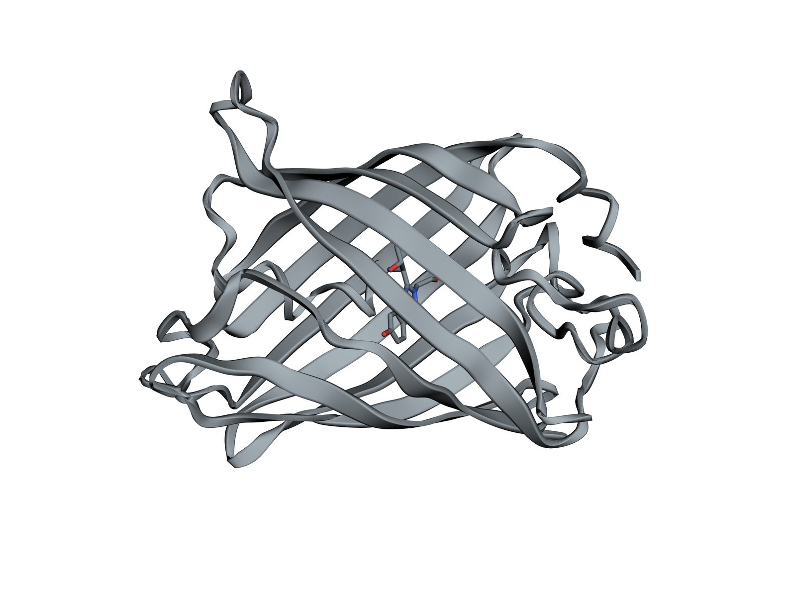
The jellyfish protein that glows green: a barrel of eleven beta strands around a chromophore it makes from three of its own amino acids.
Green fluorescent protein comes from the jellyfish Aequorea victoria. Joined to another protein by genetic engineering, it lets researchers watch where that protein goes in a living cell, because it needs nothing but oxygen to make its own light-emitting group. Its discovery and development won the 2008 Nobel Prize in Chemistry. This structure, one of the first two solved, is of the brighter S65T variant.
Also called GFP.
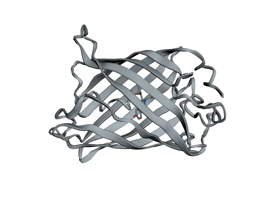
View 1 of 6
A barrel with a light inside
GFP is a chain of 238 amino acids folded into a cylinder. The light-emitting group sits at the very centre, so the barrel around it decides how brightly, and in what colour, it glows.
Use ← and → to move between views. Drag the molecule to turn it; on a touch screen, press Turn it first.
View 1 of 6: A barrel with a light inside
Download the pictureDownload the atoms (PDB)Checked against its sources on 25 October 2026.
View by view
The Green Fluorescent Protein structure, explained
01A barrel with a light inside
GFP is a chain of 238 amino acids folded into a cylinder. The light-emitting group sits at the very centre, so the barrel around it decides how brightly, and in what colour, it glows.
02Eleven strands round a can
Eleven beta strands run up and down, side by side, and close into a barrel. The ends are capped by short loops. The tight barrel makes the folded protein very stable and helps explain why its glow is so hard to quench.
03A strand up the middle
One stretch of the chain threads up the inside of the barrel. Three of its amino acids, 65, 66 and 67, are where the chromophore forms.
04A chromophore it makes itself
After the chain folds, the backbone around amino acids 65 to 67 closes into a ring, loses water and reacts with oxygen. No other enzyme or cofactor is needed, which is why GFP glows when its gene is put into almost any cell.
05The pocket sets the colour
The amino acids around the chromophore tune it. This S65T variant absorbs blue light, near 488 nanometres, and gives out green near 510. Changing the pocket gave yellow variants, and changing tyrosine 66 itself gave cyan and blue ones, used to label several proteins at once.
06From jellyfish to lab
In the jellyfish, GFP takes energy from aequorin, a protein that glows blue when calcium binds it, and gives it out as green light. In the lab, its gene joined to another protein's gene makes a glowing tag on that protein.
Parts
What each part does
- Beta barrel
- Eleven beta strands that wrap round into a closed can about 4 nanometres tall and 2.4 nanometres wide. It shields the chromophore from the surrounding water and from small molecules such as oxygen and ions.
- Central strand
- The stretch of chain that runs up the middle of the barrel, partly helical, carrying the chromophore at its centre.
- Chromophore
- The light-emitting group: two linked rings formed from amino acids 65, 66 and 67 when the chain folds. Here 65 is threonine, the S65T change that makes the protein brighter.
- Arginine 96 and glutamate 222
- Two amino acids beside the chromophore that help it form: glutamate 222 takes protons from the chain, and arginine 96 steadies the charge as the ring closes.
- Histidine 148 and threonine 203
- Two of the amino acids that hold the chromophore's tyrosine hydroxyl in place by hydrogen bonds. Changes here, as in yellow fluorescent protein, shift the colour.
The structure
Where these atoms come from
- Structure
- PDB 1EMA
- What it is
- Green fluorescent protein, S65T variant
- Organism
- Aequorea victoria
- Method
- X-ray diffraction, 1.9 Å resolution
- Deposited
- 1996
- Atoms shown
- 1,771, without water or hydrogens
Coordinates from the Protein Data Bank, free of all copyright (CC0). Water and hydrogens are left out, and the molecule is turned to face you; every atom is where the experiment placed it.
Dictionary
The terms to know
Practise it
The quizzes and cards that fit
- Biotechnology quizzes
- Bioethics & Regulatory Affairs10 questions
- Restriction Enzymes & DNA Ligation15 questions
- Plant Biotechnology & GMOs10 questions
- Cell Biology quizzes
- Characteristics of Life15 questions
- Lab Safety15 questions
- Cell Theory15 questions
- Molecular Biology quizzes
- Epigenetics & Chromatin Remodeling20 questions
- DNA Replication Machinery15 questions
- Recombinant DNA Technology15 questions
- Biotechnology flashcards
The subject's terms as a ready-made deck, free to flip through.
Next to it
More molecules
Sources
Where the facts come from
- RCSB Protein Data Bank: 1EMA, green fluorescent protein from Aequorea victoria
- Ormö and others, Crystal structure of the Aequorea victoria green fluorescent protein, Science 1996
- PDB-101 Molecule of the Month: Green Fluorescent Protein (GFP)
- Alberts and others, Molecular Biology of the Cell, 4th edition: Visualizing Molecules in Living Cells (NCBI Bookshelf)
- Day and Davidson, The fluorescent protein palette: tools for cellular imaging, Chem Soc Rev 2009 (PMC)
- Sniegowski and others, Base catalysis of chromophore formation in Arg96 and Glu222 variants of green fluorescent protein, J Biol Chem 2005
Written by biofideX from these sources. Drawn with 3Dmol.js (Rego and Koes, Bioinformatics 2015).