Potassium Channel 3D Structure
A pore in the cell membrane that lets potassium ions through in single file, about ten thousand times more readily than sodium.
Potassium channels set the resting voltage of cells and bring a nerve's action potential back down. This one, KcsA from the bacterium Streptomyces lividans, was the first ion channel whose structure was solved in atomic detail, by Roderick MacKinnon's group in 1998, work that shared the 2003 Nobel Prize in Chemistry. Its pore is built the same way as the pores of the channels in human nerve and heart.
Also called KcsA or K⁺ channel.
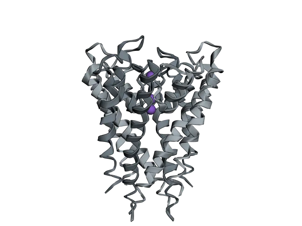
View 1 of 6
Four subunits round a pore
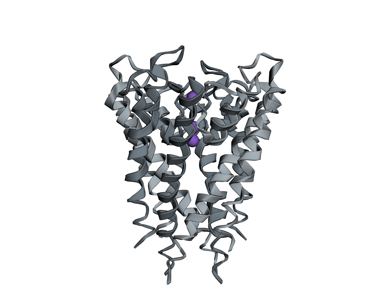
KcsA is four identical subunits round a pore down the middle. Seen from the side, as here, with the outside of the cell at the top, it is an upside-down cone: wide at the top, where the filter is, and narrow at the bottom, where the inner helices cross.
Use ← and → to move between views. Drag the molecule to turn it; on a touch screen, press Turn it first.
View 1 of 6: Four subunits round a pore
Download the pictureDownload the atoms (PDB)Checked against its sources on 25 October 2026.
View by view
The Potassium Channel structure, explained
01Four subunits round a pore
KcsA is four identical subunits round a pore down the middle. Seen from the side, as here, with the outside of the cell at the top, it is an upside-down cone: wide at the top, where the filter is, and narrow at the bottom, where the inner helices cross.
02One subunit
Each subunit crosses the membrane as two long helices, an outer one against the membrane's fats and an inner one lining the pore. Between them, at the top, a short pore helix leads into a loop that makes one quarter of the filter.
03Outer and inner helices
The four outer helices face the membrane; the four inner helices line the pore and cross near the bottom. In channels that open and close, the inner helices move apart to open this gate.
04The selectivity filter
At the top of the pore, the filter is lined by backbone carbonyl oxygens of TVGYG from all four subunits. A potassium ion entering it gives up the water around it, and the oxygens take the water's place, spaced just as the water was.
05Why sodium stays out
A sodium ion is smaller than potassium. The filter's oxygens are held too far apart to fit snugly round it, so giving up its water costs sodium more energy than the filter returns. Potassium passes about ten thousand times more readily, yet still close to the rate at which it can diffuse.
06Ions in single file
Three potassium ions sit in a line in this crystal. Ions in the filter repel each other, so each new ion entering from one end helps push the next one out of the other end, which keeps the flow fast.
Parts
What each part does
- Subunit
- One of four identical chains. Each crosses the membrane twice, as an outer and an inner helix, with a short pore helix and a loop between them at the top.
- Four subunits
- The four chains stand round a common axis, and the pore runs down the middle, where they meet.
- Outer helices
- The helix of each subunit that faces the membrane's fats. Four of them hold the channel in the membrane.
- Inner helices
- The helix of each subunit that lines the lower part of the pore. Where the four cross near the inside of the cell, they form the gate that closes the channel.
- Pore helices
- A short helix in each subunit, pointing at the centre of the channel. The negative ends of the four helices help draw a positive ion into the cavity below the filter.
- Selectivity filter
- Five amino acids of each subunit, threonine, valine, glycine, tyrosine, glycine (TVGYG), that line the narrowest part of the pore with their backbone oxygens.
- Potassium ions
- Three potassium ions placed in this model, all inside the selectivity filter, from near its outer end down to the threonines at its inner end. In a working channel they move through in single file.
The structure
Where these atoms come from
- Structure
- PDB 1BL8
- What it is
- Potassium channel KcsA
- Organism
- Streptomyces lividans
- Method
- X-ray diffraction, 3.2 Å resolution
- Deposited
- 1998
- Atoms shown
- 2,823, without water or hydrogens
Coordinates from the Protein Data Bank, free of all copyright (CC0). Water and hydrogens are left out, and the molecule is turned to face you; every atom is where the experiment placed it.
Dictionary
The terms to know
Practise it
The quizzes and cards that fit
- Neuroscience quizzes
- Reflex Arc15 questions
- Spinal Cord15 questions
- Cognitive Neuroscience: Language & Lateralization15 questions
- Physiology quizzes
- Exercise Physiology15 questions
- Puberty15 questions
- Blood Clotting15 questions
- Biochemistry quizzes
- Photosynthesis and Cellular Respiration15 questions
- Molarity and Dilutions15 questions
- ATP and Cellular Energy15 questions
- Neuroscience flashcards
The subject's terms as a ready-made deck, free to flip through.
See it at work
Diagrams it belongs to
Next to it
More molecules
Sources
Where the facts come from
- RCSB Protein Data Bank: 1BL8, potassium channel (KcsA) from Streptomyces lividans
- Doyle and others, The structure of the potassium channel: molecular basis of K⁺ conduction and selectivity, Science 1998
- PDB-101 Molecule of the Month: Potassium Channels
- Alberts and others, Molecular Biology of the Cell, 4th edition: Ion Channels and the Electrical Properties of Membranes (NCBI Bookshelf)
Written by biofideX from these sources. Drawn with 3Dmol.js (Rego and Koes, Bioinformatics 2015).