Collagen Triple Helix 3D Structure
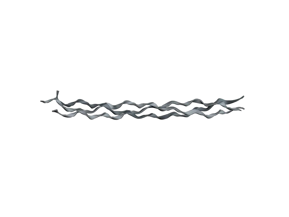
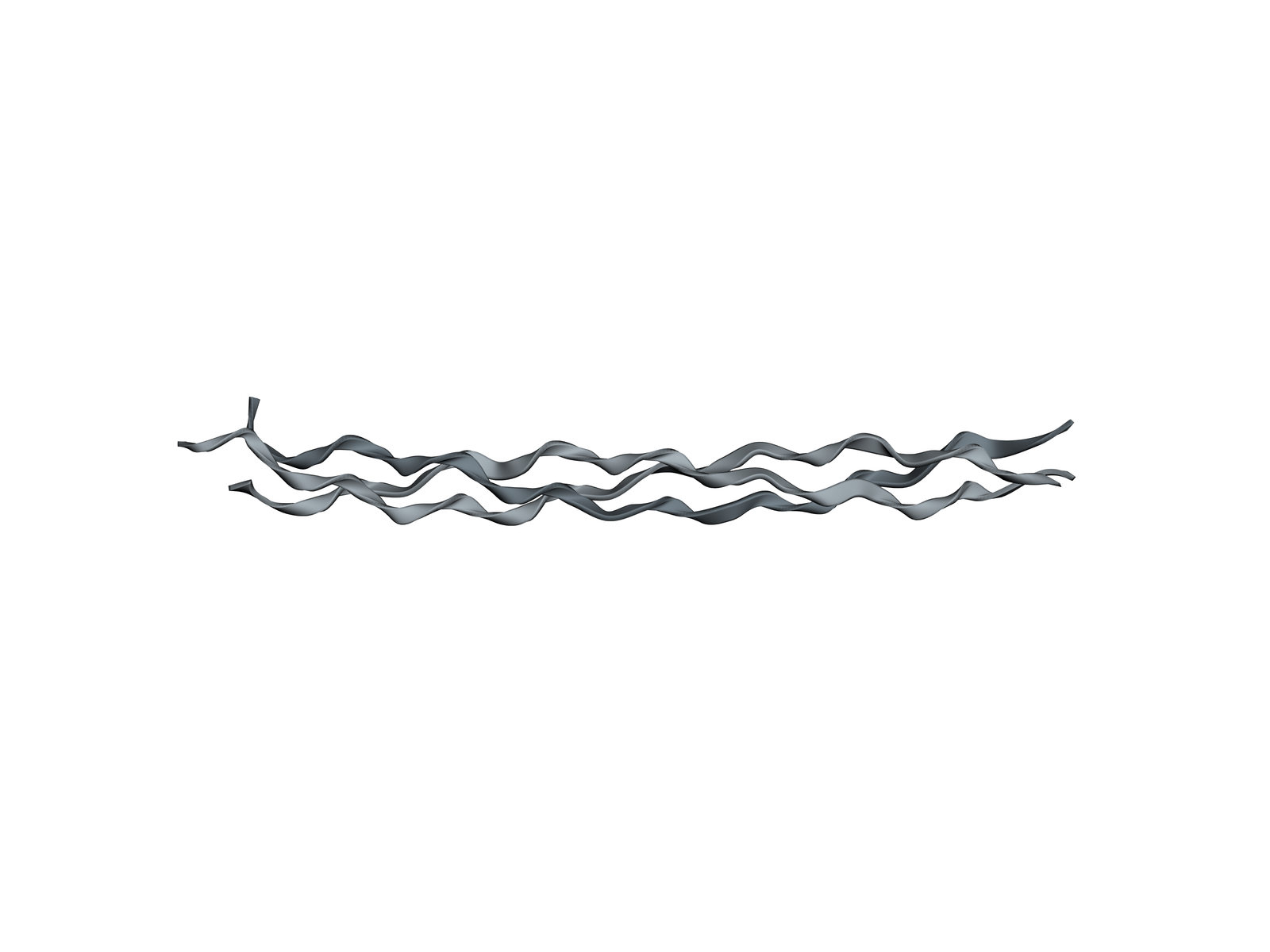
The most abundant protein in mammals: three chains wound into a rope-like triple helix, with glycine at every third position.
Collagen gives skin, tendon, bone and cartilage their strength. Its molecule is a long, thin rope of three chains, each a repeat of glycine, X, Y, where X is often proline and Y often hydroxyproline. A full collagen molecule is about 300 nanometres long; this crystal holds a short stretch, a peptide built to show how the triple helix is made.
Also called Collagen or Tropocollagen.
View 1 of 6
Three chains, one rope
The collagen molecule is a triple helix: three chains wound round each other like the strands of a rope. A full molecule is about 1.5 nanometres thick and 300 nanometres long; this piece is nearly 9 nanometres long.
Use ← and → to move between views. Drag the molecule to turn it; on a touch screen, press Turn it first.
View 1 of 6: Three chains, one rope
Download the pictureDownload the atoms (PDB)Checked against its sources on 25 October 2026.
View by view
The Collagen Triple Helix structure, explained
01Three chains, one rope
The collagen molecule is a triple helix: three chains wound round each other like the strands of a rope. A full molecule is about 1.5 nanometres thick and 300 nanometres long; this piece is nearly 9 nanometres long.
02One chain of three
Each chain is not an alpha helix but a looser, left-handed helix with about three amino acids per turn. It is stable only as part of the triple helix, held by hydrogen bonds to the other two chains.
03Glycine at every third place
The sequence repeats glycine, X, Y. Glycine sits at the centre of the triple helix, where there is no room for a larger side chain. In type I collagen, a mutation that replaces one glycine with anything bigger untwists the helix locally, and is a common cause of osteogenesis imperfecta, brittle bone disease.
04Proline and hydroxyproline
Proline and hydroxyproline make up a large share of collagen. Their rings fix the twist of each chain, and the hydroxyl of hydroxyproline adds stability, so the helix holds together at body temperature.
05Why scurvy weakens tissues
Prolyl hydroxylase, the enzyme that turns proline into hydroxyproline inside the cell, needs vitamin C. Without it, collagen chains are poorly hydroxylated and form weaker triple helices, and the gums, skin and blood vessels it holds together start to fail: scurvy.
06A real collagen sequence
The middle of this peptide is a stretch of human type III collagen, with no proline or hydroxyproline in it. The study showed that the triple helix is wound slightly differently there, so its shape follows its sequence.
Parts
What each part does
- Chain 1
- One of the three chains, about 30 amino acids here. Within the triple helix each chain twists into a narrow left-handed helix, with about three amino acids per turn.
- Chain 2
- The second chain, the same sequence as the first. In the helix it sits one amino acid along from its neighbour, so the glycines of the three chains are staggered.
- Chain 3
- The third chain. The three together twist round a common axis into a right-handed superhelix, the triple helix.
- Glycine
- Every third amino acid. Glycine has only a hydrogen for a side chain, small enough to fit at the crowded centre of the triple helix, where the three chains touch.
- Proline
- Often in the X position of glycine, X, Y. Its ring holds the chain in the twisted shape the triple helix needs.
- Hydroxyproline
- Proline with a hydroxyl added after the chain is made, by prolyl hydroxylase, an enzyme that needs vitamin C. It is usually in the Y position and makes the triple helix more stable.
- Stretch of type III collagen
- ITGARGLAG, nine amino acids of human type III collagen. With no proline or hydroxyproline in it, the helix here is wound a little differently, which the study set out to show.
The structure
Where these atoms come from
- Structure
- PDB 1BKV
- What it is
- Collagen-like peptide with a region of human type III collagen
- Organism
- Synthetic, with a sequence from Homo sapiens
- Method
- X-ray diffraction, 2 Å resolution
- Deposited
- 1998
- Atoms shown
- 563, without water or hydrogens
Coordinates from the Protein Data Bank, free of all copyright (CC0). Water and hydrogens are left out, and the molecule is turned to face you; every atom is where the experiment placed it.
Dictionary
The terms to know
Practise it
The quizzes and cards that fit
- Biochemistry quizzes
- Photosynthesis and Cellular Respiration15 questions
- Molarity and Dilutions15 questions
- ATP and Cellular Energy15 questions
- Cell Biology quizzes
- Characteristics of Life15 questions
- Lab Safety15 questions
- Cell Theory15 questions
- Human Anatomy quizzes
- Shoulder and Rotator Cuff15 questions
- Knee Joint15 questions
- Arteries and Veins15 questions
- Biochemistry flashcards
The subject's terms as a ready-made deck, free to flip through.
Next to it
More molecules
Sources
Where the facts come from
- RCSB Protein Data Bank: 1BKV, collagen
- Kramer and others, Sequence dependent conformational variations of collagen triple-helical structure, Nat Struct Biol 1999
- Bella and others, Crystal and molecular structure of a collagen-like peptide at 1.9 Å resolution, Science 1994
- PDB-101 Molecule of the Month: Collagen
- Alberts and others, Molecular Biology of the Cell, 4th edition: The Extracellular Matrix of Animals (NCBI Bookshelf)
- StatPearls: Biochemistry, Collagen Synthesis (NCBI Bookshelf)
Written by biofideX from these sources. Drawn with 3Dmol.js (Rego and Koes, Bioinformatics 2015).