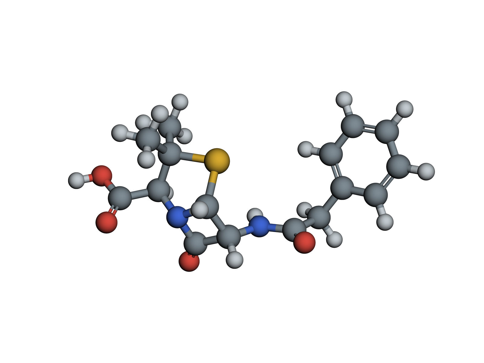
Penicillin G 3D Structure
A natural penicillin still given as a medicine: a strained four-membered beta-lactam ring that jams the enzymes bacteria use to build their cell walls.
Alexander Fleming noticed in 1928 that a Penicillium mould killed the bacteria around it; Howard Florey, Ernst Chain and their team turned its product into a medicine in the early 1940s. Penicillin G, or benzylpenicillin, is still given by injection for infections such as syphilis and streptococcal disease. Every penicillin shares its core; they differ in the side chain.
Also called Benzylpenicillin or Penicillin.
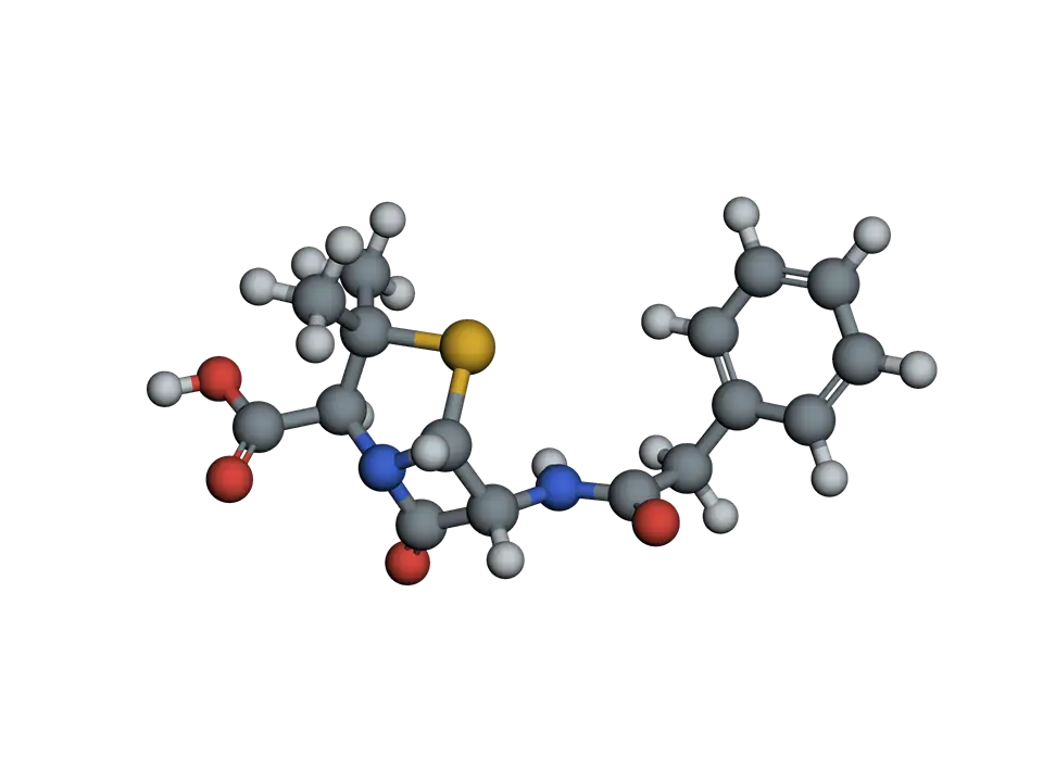
View 1 of 6
A small ring that does the work
Penicillin G is built of two fused rings, a carboxylic acid and a side chain. The square ring at its heart, the beta-lactam, is what all penicillins, cephalosporins and carbapenems share.
Use ← and → to move between views. Drag the molecule to turn it; on a touch screen, press Turn it first.
View 1 of 6: A small ring that does the work
Download the pictureDownload the atoms (SDF)Checked against its sources on 25 October 2026.
View by view
The Penicillin G structure, explained
01A small ring that does the work
Penicillin G is built of two fused rings, a carboxylic acid and a side chain. The square ring at its heart, the beta-lactam, is what all penicillins, cephalosporins and carbapenems share.
02A strained four-membered ring
The beta-lactam ring forces its bonds to angles near 90 degrees, and its nitrogen is held at the fold between two rings, so the amide is less stable than usual. Its carbonyl carbon is ready to react with the first strong nucleophile it meets.
03Jamming the wall builders
Bacteria build their cell walls of peptidoglycan, cross-linked by transpeptidases, also called penicillin-binding proteins. Penicillin looks enough like the D-alanyl-D-alanine end of the short peptides being cross-linked to enter the active site, where a serine attacks the beta-lactam and is left bound to penicillin, blocked.
04Why the bacterium bursts
Without new cross-links, the wall of a growing bacterium weakens while the cell keeps making other parts and its own enzymes keep cutting the old wall. Water pressure inside then bursts it. Human cells have no peptidoglycan, which is why penicillin is so safe for most people.
05How bacteria fight back
Many bacteria make beta-lactamases, enzymes that open the beta-lactam ring before it reaches its target. Others change their penicillin-binding proteins. Drugs such as clavulanic acid, given with amoxicillin, block beta-lactamases and protect the penicillin.
06The part that varies
Changing the side chain changes which bacteria a penicillin reaches and how it survives. Penicillin G is broken down by stomach acid, so it is injected; penicillin V and amoxicillin, with other side chains, survive the stomach and are taken by mouth.
Parts
What each part does
- Beta-lactam ring
- A four-membered ring of three carbons and a nitrogen, with a carbonyl: a cyclic amide under strain. Its carbonyl reacts readily, which is the source of penicillin's action.
- Thiazolidine ring
- A five-membered ring with a sulfur and the beta-lactam's nitrogen, carrying two methyl groups. Fused to the beta-lactam it makes the penam core of all penicillins.
- Carboxylic acid
- A COOH group on the thiazolidine ring. It is charged in the body and helps penicillin sit in the active site of its target enzymes.
- Benzyl side chain
- A benzyl group joined through an amide. Penicillins differ only here: amoxicillin and ampicillin add an amino group, which widens the range of bacteria they reach and lets them be taken by mouth.
The structure
Where these atoms come from
- Compound
- PubChem CID 5904
- Formula
- C₁₆H₁₈N₂O₄S
- Molar mass
- 334.4 g/mol
- Atoms shown
- 41, hydrogens included
- IUPAC name
- (2S,5R,6R)-3,3-dimethyl-7-oxo-6-[(2-phenylacetyl)amino]-4-thia-1-azabicyclo[3.2.0]heptane-2-carboxylic acid
- InChIKey
- JGSARLDLIJGVTE-MBNYWOFBSA-N
Coordinates from PubChem's computed 3D model, in the public domain. The model is one low-energy shape of the molecule; in water it flexes and turns about its single bonds.
Practise it
The quizzes and cards that fit
- Pharmacology quizzes
- Medication Administration15 questions
- Psychiatric Medications15 questions
- Common Drug Classes15 questions
- Microbiology quizzes
- Gut Microbiome15 questions
- Tuberculosis15 questions
- Protists15 questions
- Pharmacology flashcards
The subject's terms as a ready-made deck, free to flip through.
- benzylpenicillin drug card
A natural penicillin given by injection for streptococcal, pneumococcal and meningococcal infections, syphilis and endocarditis.
Next to it
More molecules
Sources
Where the facts come from
- PubChem: Penicillin G (CID 5904)
- StatPearls: Penicillin (NCBI Bookshelf)
- American Chemical Society, National Historic Chemical Landmarks: Discovery and Development of Penicillin
Written by biofideX from these sources. Drawn with 3Dmol.js (Rego and Koes, Bioinformatics 2015).