Transfer RNA 3D Structure
The adaptor of protein synthesis: an L-shaped RNA of 76 nucleotides that reads a codon at one end and carries an amino acid at the other.
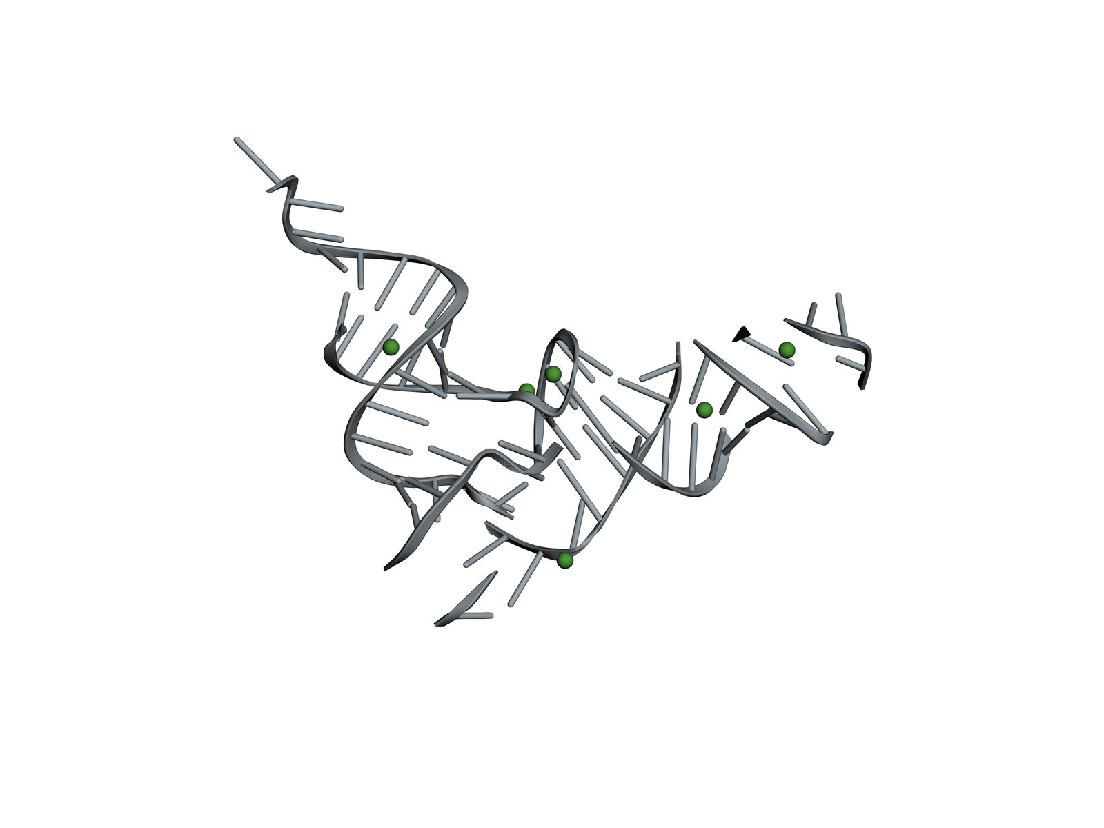
A transfer RNA links the genetic code to amino acids. Its anticodon pairs with a codon of messenger RNA in the ribosome, and its other end carries the amino acid that codon stands for. Drawn flat, its four stems make a cloverleaf; in three dimensions they fold into an L. This is the tRNA for phenylalanine from baker's yeast, the first tRNA whose structure was solved, in 1974.
Also called tRNA or tRNA for phenylalanine.
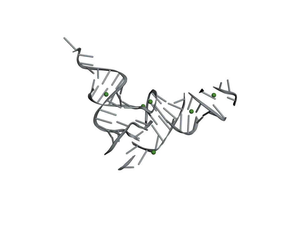
View 1 of 6
An RNA folded into an L
The tRNA's single chain of 76 nucleotides pairs with itself into four short stems, then folds so that pairs of stems stack into two arms at right angles. The anticodon and the amino acid end up about 7 nanometres apart, at the two ends of the L.
Use ← and → to move between views. Drag the molecule to turn it; on a touch screen, press Turn it first.
View 1 of 6: An RNA folded into an L
Download the pictureDownload the atoms (PDB)Checked against its sources on 25 October 2026.
View by view
The Transfer RNA structure, explained
01An RNA folded into an L
The tRNA's single chain of 76 nucleotides pairs with itself into four short stems, then folds so that pairs of stems stack into two arms at right angles. The anticodon and the amino acid end up about 7 nanometres apart, at the two ends of the L.
02Where the amino acid goes
The acceptor stem ends in four unpaired nucleotides, the last three always CCA. An enzyme matched to this tRNA, phenylalanyl-tRNA synthetase, joins phenylalanine to the final A, so the tRNA is charged.
03Where the code is read
At the other end of the L, the anticodon loop holds three unpaired bases, here Gm, A and A. In the ribosome they pair with a codon of the messenger RNA, UUC or UUU, both codons for phenylalanine.
04Four arms of a cloverleaf
Drawn flat, the tRNA is a cloverleaf of four stems: the acceptor stem, the D arm, the anticodon arm and the TΨC arm. In three dimensions the acceptor stem stacks on the TΨC arm, and the D arm on the anticodon arm.
05Held at the corner
The loops of the D and TΨC arms meet at the corner of the L, where bases from each pair and stack with each other. These tertiary contacts, with magnesium ions, hold the two arms at right angles.
06Fourteen modified nucleotides
Enzymes change many of a tRNA's bases after it is made. In this one, wybutosine sits just beside the anticodon, where it steadies codon pairing, and the modifications of the D and TΨC loops help fold the corner.
Parts
What each part does
- Acceptor stem
- The first seven nucleotides paired with seven near the far end: a short double helix that ends in the single-stranded tail where the amino acid is attached.
- CCA end
- The last three nucleotides, always C, C, A. The amino acid is joined to the sugar of the final A by an aminoacyl-tRNA synthetase.
- D arm
- A stem and loop named for the dihydrouridines in its loop. It folds against the T arm at the corner of the L.
- Anticodon arm
- A stem and a loop of seven nucleotides at the bottom of the L, which reaches into the ribosome to pair with messenger RNA.
- Anticodon
- Gm, A, A (5′ to 3′): three bases that pair with the codons UUC and UUU for phenylalanine, the second through a wobble pair at the third position.
- Variable loop
- A short loop between the anticodon arm and the T arm. Its length differs most from one tRNA to another.
- TΨC arm
- A stem and loop named for thymine, pseudouridine and cytosine in its loop. It stacks on the acceptor stem to make one long arm of the L.
- Modified nucleotides
- Fourteen of the 76 nucleotides are chemically changed after the RNA is made: methylated bases and sugars, dihydrouridine, pseudouridine and wybutosine beside the anticodon. They help the tRNA fold and read codons accurately.
- Magnesium ions
- Positive ions bound where the folded RNA brings its negative phosphates close together. They help hold the L shape.
The structure
Where these atoms come from
- Structure
- PDB 1EHZ
- What it is
- Yeast tRNA for phenylalanine
- Organism
- Saccharomyces cerevisiae
- Method
- X-ray diffraction, 1.93 Å resolution
- Deposited
- 2000
- Atoms shown
- 1,658, without water or hydrogens
Coordinates from the Protein Data Bank, free of all copyright (CC0). Water and hydrogens are left out, and the molecule is turned to face you; every atom is where the experiment placed it.
Dictionary
The terms to know
Practise it
The quizzes and cards that fit
- Molecular Biology quizzes
- Epigenetics & Chromatin Remodeling20 questions
- DNA Replication Machinery15 questions
- Recombinant DNA Technology15 questions
- Biochemistry quizzes
- Photosynthesis and Cellular Respiration15 questions
- Molarity and Dilutions15 questions
- ATP and Cellular Energy15 questions
- Genetics quizzes
- Incomplete Dominance and Codominance15 questions
- Pedigree Analysis15 questions
- Blood Types15 questions
- Molecular Biology flashcards
The subject's terms as a ready-made deck, free to flip through.
Next to it
More molecules
Sources
Where the facts come from
- RCSB Protein Data Bank: 1EHZ, the crystal structure of yeast phenylalanine tRNA at 1.93 Å resolution
- Shi and Moore, The crystal structure of yeast phenylalanine tRNA at 1.93 Å resolution: a classic structure revisited, RNA 2000
- PDB-101 Molecule of the Month: Transfer RNA
- Alberts and others, Molecular Biology of the Cell, 4th edition: From RNA to Protein (NCBI Bookshelf)
Written by biofideX from these sources. Drawn with 3Dmol.js (Rego and Koes, Bioinformatics 2015).