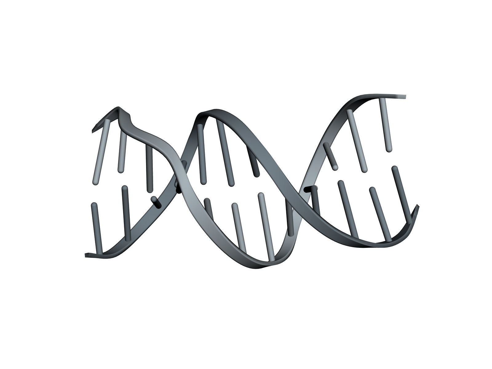
DNA Double Helix 3D Structure
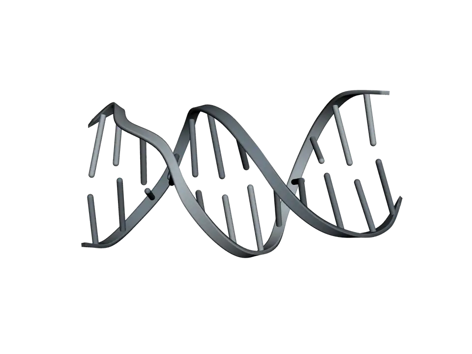
Two strands of DNA wound round each other, held together by base pairs: twelve of them here, about one full turn of the double helix.
DNA keeps its information in the order of four bases along a sugar and phosphate backbone. Two strands run in opposite directions and pair base to base, A with T and G with C, so each strand carries what is needed to rebuild the other. This crystal structure, first reported in 1980 and refined in 1981, was the first to show a whole turn of B-DNA, the form found in cells.
Also called B-DNA or Deoxyribonucleic acid.
View 1 of 7
A double helix of two strands
Two strands of DNA wind round each other in a right-handed helix about 2 nanometres across. The backbones run along the outside and the bases point inwards, where each pairs with a base on the other strand.
Use ← and → to move between views. Drag the molecule to turn it; on a touch screen, press Turn it first.
View 1 of 7: A double helix of two strands
Download the pictureDownload the atoms (PDB)Checked against its sources on 25 October 2026.
View by view
The DNA Double Helix structure, explained
01A double helix of two strands
Two strands of DNA wind round each other in a right-handed helix about 2 nanometres across. The backbones run along the outside and the bases point inwards, where each pairs with a base on the other strand.
02Two strands, running opposite ways
Each strand has a direction, from the 5′ end of its sugar chain to the 3′ end. The two run in opposite directions, so the 5′ end of one strand lies beside the 3′ end of the other. This is what antiparallel means.
03The backbone on the outside
Each sugar is joined to the next through a phosphate, by phosphodiester bonds. With the phosphates on the outside, facing the water, DNA is a strongly negatively charged molecule, which is why it moves towards the positive pole in gel electrophoresis.
04A pairs with T
Inside the helix each base pairs with one on the other strand. Adenine always pairs with thymine, through two hydrogen bonds, because only that pair has the right shape and the right donors and acceptors.
05G pairs with C
Guanine pairs with cytosine through three hydrogen bonds. Every pair, A-T or G-C, is a larger purine opposite a smaller pyrimidine, so every rung is the same width and the helix stays even.
06Stacked like coins
The flat base pairs stack about 0.34 nanometres apart, about ten to a turn. Stacking, more than the hydrogen bonds between the bases, is what holds the helix together. Twelve pairs make a little more than one full turn, as here.
07A sequence an enzyme can read
The middle six bases, GAATTC, read the same on both strands. This is the site of EcoRI, a restriction enzyme that recognises this sequence by touching the edges of the bases in the major groove, then cuts it.
Parts
What each part does
- Strand 1
- Twelve nucleotides, CGCGAATTCGCG, read from its 5′ end to its 3′ end. Each nucleotide is a phosphate, a deoxyribose sugar and a base.
- Strand 2
- The same twelve bases, running the other way. Because the sequence reads the same on both strands, the two strands are identical: a palindrome.
- Sugar and phosphate backbone
- Deoxyribose sugars joined through phosphates by phosphodiester bonds. It runs along the outside of the helix, and its phosphates carry negative charges in water.
- Bases
- Adenine, thymine, guanine and cytosine, flat rings that stack on top of each other inside the helix. Their order along a strand is the genetic information.
- A-T base pairs
- Adenine pairs with thymine through two hydrogen bonds. The four in the middle of this piece, AATT on each strand, make the helix's narrowest stretch of minor groove.
- G-C base pairs
- Guanine pairs with cytosine through three hydrogen bonds. G-C rich DNA holds its two strands together more tightly than A-T rich DNA, partly from the extra bond and partly because G-C pairs stack more strongly on their neighbours.
- EcoRI site
- GAATTC on both strands: the sequence the restriction enzyme EcoRI recognises and cuts, between the G and the first A of each strand.
The structure
Where these atoms come from
- Structure
- PDB 1BNA
- What it is
- B-DNA dodecamer, 5′ CGCGAATTCGCG 3′
- Organism
- Synthetic
- Method
- X-ray diffraction, 1.9 Å resolution
- Deposited
- 1981
- Atoms shown
- 486, without water or hydrogens
Coordinates from the Protein Data Bank, free of all copyright (CC0). Water and hydrogens are left out, and the molecule is turned to face you; every atom is where the experiment placed it.
Dictionary
The terms to know
Practise it
The quizzes and cards that fit
- Molecular Biology quizzes
- Epigenetics & Chromatin Remodeling20 questions
- DNA Replication Machinery15 questions
- Recombinant DNA Technology15 questions
- Genetics quizzes
- Incomplete Dominance and Codominance15 questions
- Pedigree Analysis15 questions
- Blood Types15 questions
- Biochemistry quizzes
- Photosynthesis and Cellular Respiration15 questions
- Molarity and Dilutions15 questions
- ATP and Cellular Energy15 questions
- Molecular Biology flashcards
The subject's terms as a ready-made deck, free to flip through.
See it at work
Diagrams it belongs to
Next to it
More molecules
Sources
Where the facts come from
- RCSB Protein Data Bank: 1BNA, Structure of a B-DNA dodecamer
- Drew and others, Structure of a B-DNA dodecamer: conformation and dynamics, Proc Natl Acad Sci U S A 1981
- Wing and others, Crystal structure analysis of a complete turn of B-DNA, Nature 1980
- Yakovchuk, Protozanova and Frank-Kamenetskii, Base-stacking and base-pairing contributions into thermal stability of the DNA double helix, Nucleic Acids Res 2006 (PMC)
- Alberts and others, Molecular Biology of the Cell, 4th edition: The Structure and Function of DNA (NCBI Bookshelf)
- PDB-101 Molecule of the Month: DNA
Written by biofideX from these sources. Drawn with 3Dmol.js (Rego and Koes, Bioinformatics 2015).