Codon Chart
All 64 mRNA codons and the amino acid each one codes for, with the start codon, the three stop codons and how to read the chart.
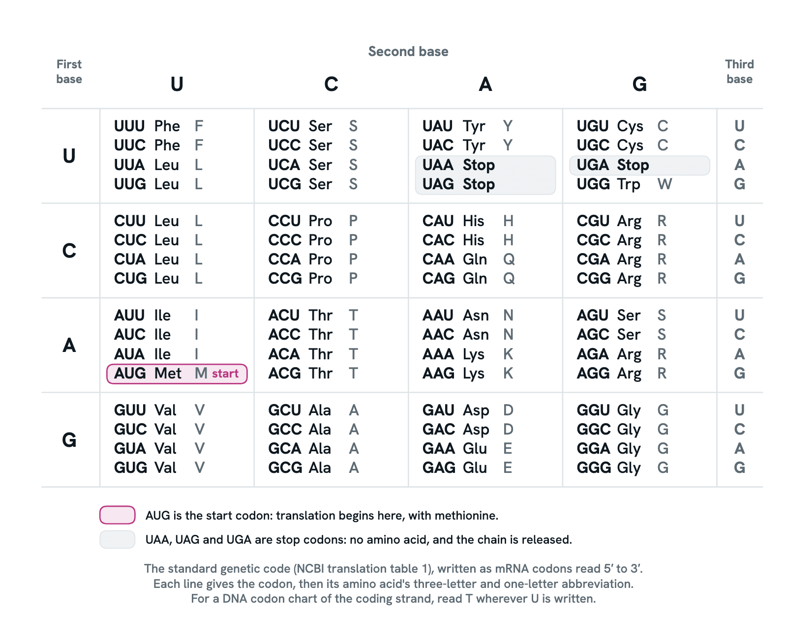
A codon chart, or codon table, is the genetic code laid out so that any codon can be looked up: find the row of its first base, the column of its second and the line of its third, and read off the amino acid. This is the standard code, written in mRNA bases, so U stands where a DNA chart would show T. Press any codon to light up every codon for the same amino acid, with what that amino acid is and does.
Also called Codon table, mRNA codon chart, Genetic code chart, Amino acid codon chart or Codon wheel.
Press any part of the diagram, or pick one here, to see what it is and what it does.
Download the chartChecked against its sources on 4 November 2026.
How to read it
How to read the codon chart
01The codon chart at a glance
The genetic code is read in codons, three bases of mRNA at a time. Four bases give 64 codons: 61 code for an amino acid and 3 say stop. The chart lays all 64 out in a grid, so any codon can be looked up by its three bases. There are only 20 amino acids, so most are coded by more than one codon.
02The first base picks the row
Start with the codon's first base, at its 5′ end, and find its row down the left side: U, C, A or G. Every codon in that row begins with it. The bases are listed in the order U, C, A, G, the order codon tables use by convention; NCBI's tables follow it too, with T written for U.
03The second base picks the column
The second base picks the column across the top. Row and column together land on one box of four codons that share their first two bases. For eight amino acids that box is all one amino acid: every codon starting GC is alanine, every codon starting GG is glycine, whatever the third base.
04The third base picks the line, so CAU is histidine
The third base picks the line within the box, using the letters at the right. So for CAU: row C, column A, line U, which reads histidine, His, H. Try it with any codon from a sequence. The translation tool linked below does the same lookup for a whole sequence at once.
05AUG, the start codon
AUG codes for methionine and is also where translation begins. In eukaryotes the ribosome scans along the mRNA from its 5′ end and usually starts at the first AUG it meets, so almost every new protein starts with methionine, which is often trimmed off later. The bases before the start codon are not translated.
06UAA, UAG and UGA, the stop codons
Three codons code for no amino acid. When one of them reaches the ribosome, no tRNA matches it; a release factor binds instead and the finished chain is set free. Their old names, from the mutations they were found in, are ochre (UAA), amber (UAG) and opal (UGA). A mutation that turns a codon into a stop is a nonsense mutation.
07Most amino acids have several codons
With 61 codons for 20 amino acids, the code is redundant, or degenerate. Leucine, serine and arginine have six codons each; most have two or four; only methionine and tryptophan have one. Codons for the same amino acid usually differ in the third base, which pairs loosely with the tRNA's anticodon: Crick's wobble. So one tRNA can read several codons.
08One base changed: silent, missense or nonsense
Because of the redundancy, a change in the third base often leaves the amino acid the same: a silent mutation. A change that swaps one amino acid for another is a missense mutation. In sickle cell disease the sixth codon of the beta-globin gene reads GUG instead of GAG, so valine replaces glutamate, and the hemoglobin sticks together into fibres.
09Nearly universal, with a few exceptions
The same chart serves bacteria, plants and people, which is why a human gene can be read by a bacterium. But there are small variations. In the mitochondria of vertebrates, UGA codes for tryptophan instead of stop, AUA for methionine instead of isoleucine, and AGA and AGG, arginine elsewhere, are listed as stop codons, though in human mitochondria they are now thought to end translation by a frameshift onto a UAG. Some ciliates and yeasts read a codon or two differently.
10mRNA chart or DNA chart
This chart is written in mRNA bases, which is what the ribosome reads. A DNA codon chart shows the same code in the coding strand, with T in place of U: AUG becomes ATG. To translate the template strand, take its reverse complement first, then read the result off the chart as DNA.
Entries
What each part of the codon chart shows
- First base (the row)
- The first base of the codon, at its 5′ end, picks the row: U, C, A or G. Read a codon from left to right, as the ribosome reads mRNA from 5′ to 3′.
- Second base (the column)
- The second base picks the column. The first two bases together often settle the amino acid on their own: every codon beginning GC is alanine, every codon beginning CC is proline.
- Third base (the line)
- The third base, at the 3′ end, picks the line within the box. It is the base that matters least: for eight amino acids any third base gives the same answer, which is why a mutation here is often silent.
- Alanine (Ala, A)
- Alanine (Ala, A). Nonpolar, with the smallest side chain after glycine. Four codons, GCU, GCC, GCA and GCG: the third base makes no difference. Made from pyruvate in one step, it carries nitrogen from muscle to the liver in the glucose-alanine cycle.
- Arginine (Arg, R)
- Arginine (Arg, R). Positively charged. Six codons: CGU, CGC, CGA, CGG, AGA and AGG. The body makes it in the urea cycle, but not always enough, so it is conditionally essential. It is also the source of nitric oxide.
- Asparagine (Asn, N)
- Asparagine (Asn, N). Polar and uncharged, the amide of aspartate. Two codons, AAU and AAC. Sugar chains are attached to proteins at asparagine in N-linked glycosylation. The first amino acid to be isolated, from asparagus juice, in 1806.
- Aspartic acid (aspartate) (Asp, D)
- Aspartic acid, or aspartate (Asp, D). Negatively charged. Two codons, GAU and GAC. It feeds the urea cycle, supplies atoms to purines and pyrimidines, and carries reducing power into mitochondria in the malate-aspartate shuttle.
- Cysteine (Cys, C)
- Cysteine (Cys, C). Polar, with a sulfur-containing thiol group. Two codons, UGU and UGC. Two cysteines can bond across a protein as a disulfide bridge, which holds insulin, antibodies and keratin in shape. Made from methionine's sulfur and serine.
- Glutamine (Gln, Q)
- Glutamine (Gln, Q). Polar and uncharged, the amide of glutamate. Two codons, CAA and CAG. The most abundant free amino acid in blood, it carries ammonia safely between tissues and donates nitrogen for making nucleotides.
- Glutamic acid (glutamate) (Glu, E)
- Glutamic acid, or glutamate (Glu, E). Negatively charged. Two codons, GAA and GAG. The hub of nitrogen metabolism, where most amino groups are exchanged, and the brain's main excitatory neurotransmitter. GAG to GUG, glutamate to valine, is the sickle cell mutation.
- Glycine (Gly, G)
- Glycine (Gly, G). The smallest amino acid: its side chain is a single hydrogen, so it is the only one without a chiral centre. Four codons, GGU, GGC, GGA and GGG. Every third residue of collagen is glycine, and it is an inhibitory neurotransmitter in the spinal cord.
- Histidine (His, H)
- Histidine (His, H). Positively charged, though its imidazole ring is only partly charged at the pH of the body, which lets it pass protons about in enzyme active sites. Two codons, CAU and CAC. Essential in the diet, and the source of histamine.
- Isoleucine (Ile, I)
- Isoleucine (Ile, I). Nonpolar and branched, essential in the diet. Three codons, AUU, AUC and AUA; in the vertebrate mitochondrial code AUA is read as methionine instead. One of the three branched-chain amino acids, with leucine and valine.
- Leucine (Leu, L)
- Leucine (Leu, L). Nonpolar and branched, essential in the diet. Six codons, as many as any amino acid has: UUA, UUG, CUU, CUC, CUA and CUG. The commonest amino acid in proteins, and a signal that amino acids are plentiful, which stimulates protein synthesis in muscle.
- Lysine (Lys, K)
- Lysine (Lys, K). Positively charged, essential in the diet. Two codons, AAA and AAG. Its side chain is where histones are acetylated and methylated to control genes, and where collagen fibres are cross-linked. The amino acid in shortest supply in cereal grains.
- Methionine (Met, M), the start codon
- Methionine (Met, M). Nonpolar, with a sulfur in its side chain, and essential in the diet. One codon, AUG, which is also the start codon: nearly every protein is begun with methionine, often removed afterwards. As S-adenosylmethionine it donates methyl groups.
- Phenylalanine (Phe, F)
- Phenylalanine (Phe, F). Aromatic and nonpolar, essential in the diet. Two codons, UUU and UUC. The body turns it into tyrosine; in phenylketonuria that enzyme is faulty or missing, so phenylalanine builds up and has to be limited in the diet.
- Proline (Pro, P)
- Proline (Pro, P). Nonpolar, and the only amino acid whose side chain loops back onto the backbone nitrogen. Four codons, CCU, CCC, CCA and CCG. The ring is stiff, so proline bends and breaks helices, and hydroxylated proline strengthens collagen.
- Serine (Ser, S)
- Serine (Ser, S). Polar, with a hydroxyl group. Six codons: UCU, UCC, UCA, UCG, AGU and AGC. Kinases attach phosphate to serine to switch proteins on and off, serine proteases cut with it, and it is the starting material for glycine, cysteine and many phospholipids.
- Threonine (Thr, T)
- Threonine (Thr, T). Polar, with a hydroxyl group, and essential in the diet. Four codons, ACU, ACC, ACA and ACG. Like serine it is a site for phosphorylation and for attaching sugars, and like isoleucine it has two chiral centres.
- Tryptophan (Trp, W)
- Tryptophan (Trp, W). Aromatic, with a large indole ring, and essential in the diet. One codon, UGG, and the rarest amino acid in proteins. The source of serotonin, melatonin and niacin. In the vertebrate mitochondrial code UGA is read as tryptophan too.
- Tyrosine (Tyr, Y)
- Tyrosine (Tyr, Y). Aromatic, with a phenol group. Two codons, UAU and UAC. Made from phenylalanine, so not essential while that is supplied. Tyrosine kinases phosphorylate it to pass signals, and it is the source of dopamine, adrenaline, thyroid hormones and melanin.
- Valine (Val, V)
- Valine (Val, V). Nonpolar and branched, essential in the diet. Four codons, GUU, GUC, GUA and GUG. In sickle cell disease a single base change, GAG to GUG, puts valine where glutamate belongs at the sixth position of the beta-globin chain.
- Stop codons (UAA, UAG, UGA)
- No tRNA carries an anticodon for a stop codon; a release factor binds in its place and the finished chain is let go. Their old names are ochre, amber and opal. UGA can also code for selenocysteine, a 21st amino acid, where the mRNA carries a special signal downstream.
- What the chart shows
- The standard genetic code, NCBI translation table 1, used by the nuclear genes of nearly every organism. It is written as mRNA codons; a DNA codon chart for the coding strand reads T wherever this one reads U. The abbreviations follow the IUPAC-IUBMB nomenclature.
As a table
The codon chart as a table
The 20 amino acids of the standard genetic code, with their abbreviations, their codons, the class of their side chain and whether the diet must supply them.
Alanine
- Abbreviations
- Ala, A
- mRNA codons
- GCU, GCC, GCA, GCG
- Side chain
- Nonpolar, aliphatic
- In the diet
- Not essential
Arginine
- Abbreviations
- Arg, R
- mRNA codons
- CGU, CGC, CGA, CGG, AGA, AGG
- Side chain
- Positively charged
- In the diet
- Conditionally essential
Asparagine
- Abbreviations
- Asn, N
- mRNA codons
- AAU, AAC
- Side chain
- Polar, uncharged
- In the diet
- Not essential
Aspartic acid (aspartate)
- Abbreviations
- Asp, D
- mRNA codons
- GAU, GAC
- Side chain
- Negatively charged
- In the diet
- Not essential
Cysteine
- Abbreviations
- Cys, C
- mRNA codons
- UGU, UGC
- Side chain
- Polar, uncharged
- In the diet
- Conditionally essential
Glutamine
- Abbreviations
- Gln, Q
- mRNA codons
- CAA, CAG
- Side chain
- Polar, uncharged
- In the diet
- Conditionally essential
Glutamic acid (glutamate)
- Abbreviations
- Glu, E
- mRNA codons
- GAA, GAG
- Side chain
- Negatively charged
- In the diet
- Not essential
Glycine
- Abbreviations
- Gly, G
- mRNA codons
- GGU, GGC, GGA, GGG
- Side chain
- Nonpolar, aliphatic
- In the diet
- Conditionally essential
Histidine
- Abbreviations
- His, H
- mRNA codons
- CAU, CAC
- Side chain
- Positively charged
- In the diet
- Essential
Isoleucine
- Abbreviations
- Ile, I
- mRNA codons
- AUU, AUC, AUA
- Side chain
- Nonpolar, aliphatic
- In the diet
- Essential
Leucine
- Abbreviations
- Leu, L
- mRNA codons
- UUA, UUG, CUU, CUC, CUA, CUG
- Side chain
- Nonpolar, aliphatic
- In the diet
- Essential
Lysine
- Abbreviations
- Lys, K
- mRNA codons
- AAA, AAG
- Side chain
- Positively charged
- In the diet
- Essential
Methionine
- Abbreviations
- Met, M
- mRNA codons
- AUG
- Side chain
- Nonpolar, aliphatic
- In the diet
- Essential
Phenylalanine
- Abbreviations
- Phe, F
- mRNA codons
- UUU, UUC
- Side chain
- Aromatic
- In the diet
- Essential
Proline
- Abbreviations
- Pro, P
- mRNA codons
- CCU, CCC, CCA, CCG
- Side chain
- Nonpolar, aliphatic
- In the diet
- Conditionally essential
Serine
- Abbreviations
- Ser, S
- mRNA codons
- UCU, UCC, UCA, UCG, AGU, AGC
- Side chain
- Polar, uncharged
- In the diet
- Not essential
Threonine
- Abbreviations
- Thr, T
- mRNA codons
- ACU, ACC, ACA, ACG
- Side chain
- Polar, uncharged
- In the diet
- Essential
Tryptophan
- Abbreviations
- Trp, W
- mRNA codons
- UGG
- Side chain
- Aromatic
- In the diet
- Essential
Tyrosine
- Abbreviations
- Tyr, Y
- mRNA codons
- UAU, UAC
- Side chain
- Aromatic
- In the diet
- Conditionally essential
Valine
- Abbreviations
- Val, V
- mRNA codons
- GUU, GUC, GUA, GUG
- Side chain
- Nonpolar, aliphatic
- In the diet
- Essential
Stop
- Abbreviations
- Stop, *
- mRNA codons
- UAA, UAG, UGA
- Side chain
- No amino acid: the chain is released
AUG, methionine's one codon, is also the start codon. Essential means the body cannot make it, so food must supply it; conditionally essential means the body usually makes enough, but not in infancy, illness or rapid growth. Side chains are classed as in Lehninger Principles of Biochemistry.
Dictionary
The terms to know
Practise it
The quizzes and cards that fit
- Molecular Biology quizzes
- Epigenetics & Chromatin Remodeling20 questions
- DNA Replication Machinery15 questions
- Recombinant DNA Technology15 questions
- Genetics quizzes
- Incomplete Dominance and Codominance15 questions
- Pedigree Analysis15 questions
- Blood Types15 questions
- Biochemistry quizzes
- Photosynthesis and Cellular Respiration15 questions
- Molarity and Dilutions15 questions
- ATP and Cellular Energy15 questions
- Molecular Biology flashcards
The subject's terms as a ready-made deck, free to flip through.
- DNA to Protein Translation
Translate DNA or RNA into protein in any of the six frames, with the standard or a mitochondrial or bacterial code, and get the protein's mass and pI.
- ORF Finder
Every open reading frame in all six frames, with its position, length and protein, from ATG or any start codon.
In 3D
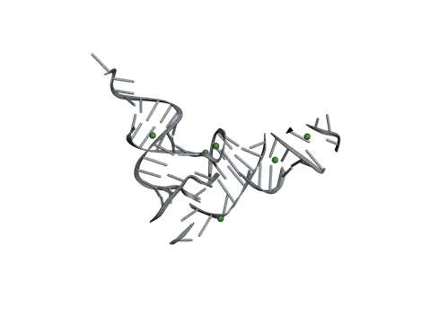
The molecules, to turn and explore
Next to it
More diagrams
Sources
Where the facts come from
- NCBI: The Genetic Codes, translation table 1 (the standard code)
- IUPAC-IUBMB Joint Commission on Biochemical Nomenclature: Nomenclature and Symbolism for Amino Acids and Peptides
- Alberts and others, Molecular Biology of the Cell, 4th edition: From RNA to Protein (NCBI Bookshelf)
- National Human Genome Research Institute: Stop Codon (Talking Glossary of Genomic and Genetic Terms)
- Crick: Codon-anticodon pairing, the wobble hypothesis, Journal of Molecular Biology 1966 (PubMed)
- Hatfield and Gladyshev: How selenium has altered our understanding of the genetic code, Molecular and Cellular Biology 2002 (PubMed)
- StatPearls: Biochemistry, Essential Amino Acids, archived chapter (NCBI Bookshelf)
- Watford and Wu: Protein, Advances in Nutrition 2018 (PubMed), on the conditionally indispensable amino acids
- MedlinePlus Genetics: HBB gene (the sickle cell variant, Glu6Val)
Drawn and written by biofideX from these sources.