Neuron Diagram
The parts of a neuron and what they do: dendrites, cell body, axon hillock, axon, myelin sheath, nodes of Ranvier, axon terminals and the synapse.
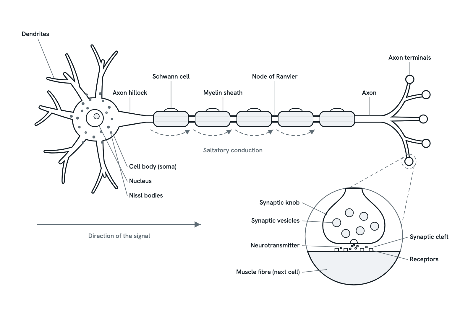
A motor neuron, the nerve cell that carries commands from the spinal cord to a muscle, laid out so its signal runs from left to right, with one synapse drawn large below. Press any part to see what it does, or take the tour. Not to scale: the axon can be about a metre long.
Also called Nerve cell or Structure of a neuron.
Press any part of the diagram, or pick one here, to see what it is and what it does.
Download the diagramChecked against its sources on 29 October 2026.
A tour
A tour of the neuron
01The neuron at a glance
A neuron is a cell built to carry signals. This one is a motor neuron, which carries commands from the spinal cord to a muscle. Like most neurons it has three main parts: dendrites that receive signals, a cell body that keeps the cell alive, and a single long axon that sends the signal on.
02Dendrites and the cell body
Dendrites branch out from the cell body and receive signals from other neurons at synapses on their surface. The cell body holds the nucleus and the Nissl bodies, clumps of rough endoplasmic reticulum where the neuron makes its proteins.
03The axon hillock: fire or not
Each incoming signal nudges the membrane potential up or down a little. The changes spread to the axon hillock and the initial segment just beyond it, where they are summed. If the total reaches threshold, about −55 mV, an action potential starts, usually in the initial segment; if not, nothing is sent. The signal is all or none.
04The axon and its myelin
The axon carries the action potential away from the cell body. Along most of its length it is wrapped in myelin: in a nerve, outside the spinal cord, each segment is a Schwann cell wound round the axon many times. The fatty layers insulate the axon, so far less charge leaks out through its membrane.
05Nodes of Ranvier: saltatory conduction
Between the segments of myelin are short bare gaps, the nodes of Ranvier, crowded with voltage-gated sodium channels. The action potential is renewed only at the nodes, so it seems to jump from one to the next: saltatory conduction, from the Latin for leap. This makes a myelinated axon conduct far faster than a bare one of the same width.
06Axon terminals
At its end the axon divides into fine branches, each ending in a swelling, the synaptic knob. Each knob meets the next cell at a synapse. For a motor neuron the next cells are skeletal muscle fibres, and one neuron's branches can reach many fibres at once.
07Across the synapse
When the action potential reaches a knob, calcium flows in and vesicles fuse with the membrane, releasing neurotransmitter into the synaptic cleft. It diffuses across and binds to receptors on the next cell. At a muscle fibre the transmitter is acetylcholine, and binding starts the electrical signal that makes the fibre contract.
08One way only
A signal passes through a neuron one way, as the arrow shows: in at the dendrites and cell body, out along the axon to the terminals. Only the knobs release neurotransmitter, and the patch of axon that has just fired is briefly refractory, so the action potential cannot turn back.
Parts
What each part of the neuron does
- Dendrites
- Short, branching processes that receive signals from other neurons, mostly at synapses on their surface, and carry them to the cell body. Their branches give a large surface for incoming signals.
- Cell body (soma)
- Holds the nucleus and most of the organelles, and makes the proteins the neuron needs. Signals from the dendrites pass through it to the axon. A motor neuron's cell body lies in the spinal cord or brainstem.
- Nucleus
- Holds the neuron's DNA. It is large and round, with a clear nucleolus, a sign of a cell that makes a great deal of protein. Mature neurons do not divide, so most must last a lifetime.
- Nissl bodies
- Clumps of rough endoplasmic reticulum and ribosomes in the cell body and the bases of the dendrites, where the neuron makes its proteins, including those it needs for its neurotransmitter. There are none in the axon hillock or the axon.
- Axon hillock
- The cone where the axon leaves the cell body. Here and in the stretch just beyond it, the initial segment, the incoming signals are summed; if they reach threshold, an action potential fires, usually starting in the initial segment.
- Axon
- The single long process that carries action potentials away from the cell body. A motor neuron's axon runs out in a nerve to a muscle and can be about a metre long.
- Myelin sheath
- A fatty covering made of many layers of cell membrane wrapped round the axon, in segments. It insulates the axon, so the signal travels much faster. Myelinated axons make up the white matter of the brain and spinal cord.
- Schwann cells
- Outside the brain and spinal cord, each segment of myelin is one Schwann cell wound round the axon many times, its nucleus pressed to the outside. Inside them, cells called oligodendrocytes make the myelin instead.
- Nodes of Ranvier
- Short bare gaps between the segments of myelin, where the axon's membrane is exposed and packed with voltage-gated sodium channels. The action potential is renewed at each node in turn.
- Axon terminals
- The axon ends in fine branches, each swelling into a synaptic knob (also called a terminal bouton). Each knob meets the next cell at a synapse, where the signal is passed on by a chemical.
- Synaptic vesicles
- Tiny membrane sacs in the knob, each filled with neurotransmitter. When an action potential arrives, calcium flows into the knob and vesicles fuse with its membrane, releasing their contents by exocytosis.
- Neurotransmitter
- The chemical messenger released into the cleft. Where a motor neuron meets a skeletal muscle fibre, it is acetylcholine.
- Synaptic cleft
- The narrow gap between the knob and the next cell. The neurotransmitter crosses it by diffusion in a tiny fraction of a millisecond.
- Receptors
- Proteins in the next cell's membrane that the neurotransmitter binds to. On a muscle fibre they are ion channels that open when acetylcholine binds, starting an electrical signal in the fibre.
- Muscle fibre (next cell)
- A motor neuron passes its signal to skeletal muscle fibres, at neuromuscular junctions, and the fibres contract. Other neurons pass theirs to another neuron, a gland, or smooth or heart muscle.
Dictionary
The terms to know
Practise it
The quizzes and cards that fit
- Neuroscience quizzes
- Reflex Arc15 questions
- Spinal Cord15 questions
- Cognitive Neuroscience: Language & Lateralization15 questions
- Cell Biology quizzes
- Characteristics of Life15 questions
- Lab Safety15 questions
- Cell Theory15 questions
- Physiology quizzes
- Exercise Physiology15 questions
- Puberty15 questions
- Blood Clotting15 questions
- Neuroscience flashcards
The subject's terms as a ready-made deck, free to flip through.
Next to it
More diagrams
Sources
Where the facts come from
- NCI SEER Training Modules: Nerve Tissue
- NINDS: Brain Basics: Know Your Brain
- StatPearls: Neuroanatomy, Neurons (NCBI Bookshelf)
- StatPearls: Histology, Axon (NCBI Bookshelf)
- StatPearls: Neuroanatomy, Nodes of Ranvier (NCBI Bookshelf)
- StatPearls: Histology, Schwann Cells (NCBI Bookshelf)
- StatPearls: Physiology, Synapse (NCBI Bookshelf)
- StatPearls: Physiology, Neuromuscular Junction (NCBI Bookshelf)
- Yamada and Kuba, Structural and Functional Plasticity at the Axon Initial Segment, Front Cell Neurosci 2016 (PMC)
Drawn and written by biofideX from these sources.