Human Brain Diagram
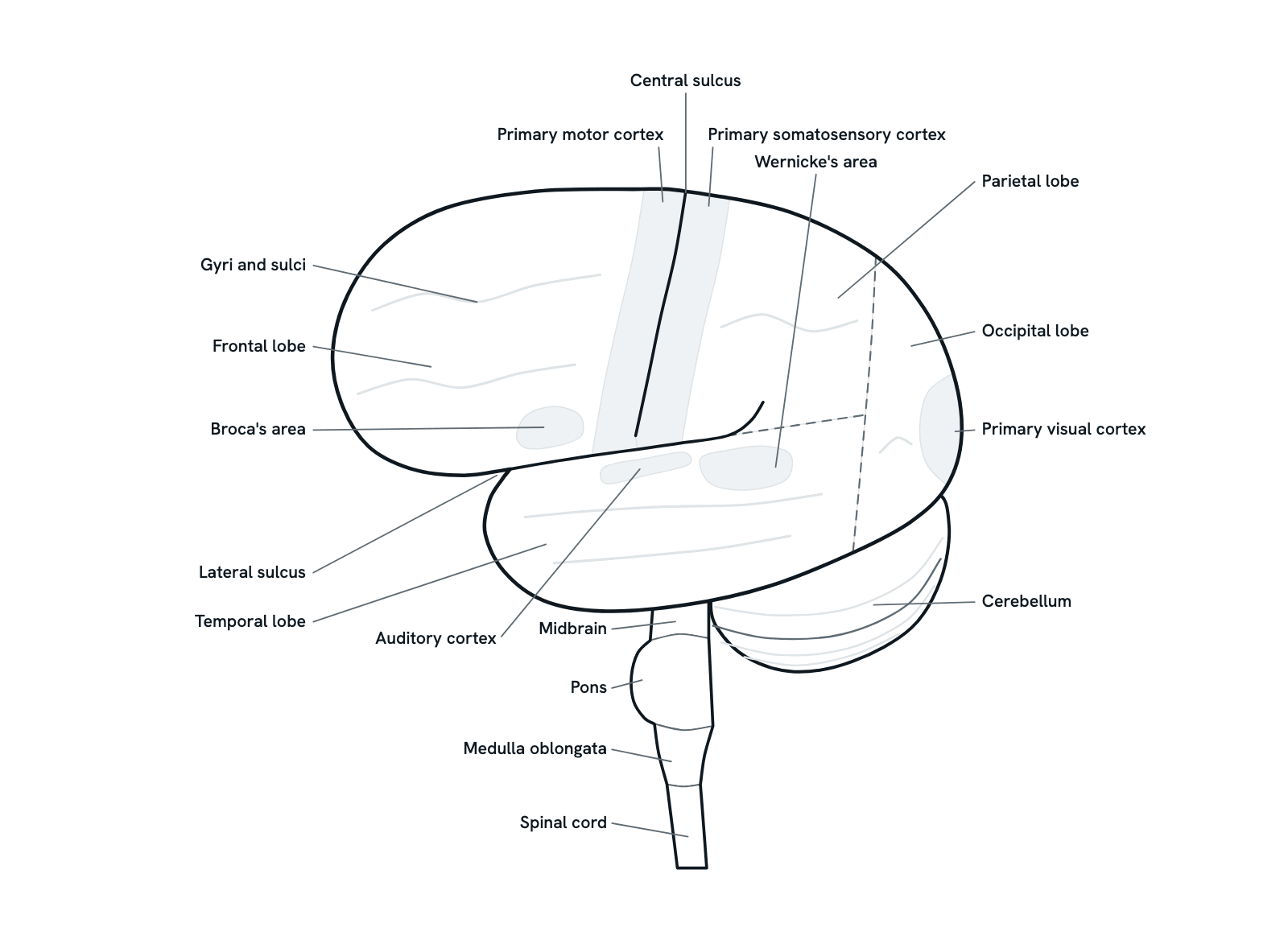
The human brain from the side: the four lobes, the cerebellum, the brainstem and the cortex areas for movement, touch, speech, hearing and sight.
The left side of the brain, front to the left: the cerebrum with its four lobes and the grooves between them, the cerebellum tucked beneath, and the brainstem running down into the spinal cord. The shaded regions are the main functional areas of the cortex. Press any part to see what it does, or take the tour. Simplified, not to scale.
Also called Parts of the brain or Lobes of the brain.
Press any part of the diagram, or pick one here, to see what it is and what it does.
Download the diagramChecked against its sources on 29 October 2026.
A tour
A tour of the human brain
01The brain at a glance
Seen from the side, three main parts of the brain show. The cerebrum, the large folded upper part, is divided into lobes. The cerebellum sits beneath its back, and the brainstem runs down from its base to become the spinal cord. An adult brain weighs about 1.4 kg (3 pounds).
02Folds and landmarks
The cerebral cortex, a thin layer of grey matter, is folded into gyri and sulci, which give it a far larger surface. Two deep grooves divide each side into lobes: the central sulcus runs down from the top, and the lateral sulcus runs back from the front, above the temporal lobe.
03The frontal lobe
The frontal lobe, the largest, lies behind the forehead, in front of the central sulcus. It plans, reasons and decides, holds one idea in mind while others are weighed, and shapes personality and behaviour. Damage here can change judgement, motivation and personality.
04The parietal lobe
The parietal lobe lies behind the central sulcus and above the lateral sulcus. Its front strip receives the body's sensations: touch, pressure, temperature, pain and position. The lobe also supports skills such as reading and arithmetic.
05Movement and touch
Either side of the central sulcus lies a strip. The primary motor cortex, in front, sends the commands for voluntary movement; the primary somatosensory cortex, behind, receives touch, pain, temperature and position. Each works for the opposite side of the body, which is mapped part by part along the strip: the leg near the top, the face low down by the lateral sulcus.
06Language: Broca's and Wernicke's areas
In most people, language depends mainly on the left hemisphere. Broca's area, in the frontal lobe, is needed to produce speech, and Wernicke's area, in the temporal lobe, to understand it. Damage to Broca's area leaves speech slow and effortful; damage to Wernicke's area leaves it fluent but with little meaning.
07Hearing and sight
The temporal lobe, below the lateral sulcus, holds the auditory cortex, which receives signals from the ears, and on its inner side structures needed to form memories. The occipital lobe at the back is given over to vision: the primary visual cortex receives signals from the eyes, each side handling the opposite half of what we see.
08The cerebellum
The cerebellum, under the occipital lobes and behind the brainstem, coordinates movement. It does not start movements but makes them smooth and accurate, keeps balance and posture, and helps learn skilled movements such as playing an instrument. Damage makes movements clumsy and unsteady.
09The brainstem and spinal cord
The brainstem links the cerebrum and cerebellum with the spinal cord. From top to bottom it is the midbrain, the pons and the medulla oblongata. It keeps breathing, heart rate, blood pressure, and sleep and waking in order, and the signals between brain and body pass through it. At the base of the skull the medulla becomes the spinal cord.
Parts
What each part of the human brain does
- Frontal lobe
- The largest lobe, behind the forehead and in front of the central sulcus. It plans, reasons and decides, holds ideas in mind while others are weighed, and shapes personality and behaviour. Its back strip is the primary motor cortex.
- Parietal lobe
- Behind the central sulcus and above the lateral sulcus. Its front strip receives touch, temperature, pain and the sense of where the body is, and the lobe as a whole supports skills such as reading and arithmetic.
- Temporal lobe
- Below the lateral sulcus, at the side of the head. It holds the auditory cortex and, usually on the left, Wernicke's area. On its inner side lie the hippocampus, needed to form new memories, and the amygdala.
- Occipital lobe
- The smallest lobe, at the back of the head, resting above the cerebellum. It processes vision. From the side its front border is not one clear groove, so it is drawn dashed here.
- Central sulcus
- The groove that runs down each side of the brain from the top, between the frontal lobe in front and the parietal lobe behind. The motor strip lies just in front of it, the touch strip just behind.
- Lateral sulcus
- Also called the Sylvian fissure: the deep groove between the temporal lobe below and the frontal and parietal lobes above. A fifth lobe, the insula, is hidden deep inside it.
- Gyri and sulci
- The cerebral cortex, the outer layer of grey matter only a few millimetres thick, is where most of the brain's processing happens. It is folded into ridges (gyri) and grooves (sulci), which fit a large surface inside the skull.
- Primary motor cortex
- A strip of the frontal lobe just in front of the central sulcus, on the precentral gyrus (Brodmann area 4). It sends the commands for voluntary movement, each part of the strip serving a part of the opposite side of the body.
- Primary somatosensory cortex
- A strip of the parietal lobe just behind the central sulcus, on the postcentral gyrus. It receives touch, pressure, temperature, pain and body position from the opposite side of the body, mapped part by part along the strip.
- Broca's area
- In the frontal lobe, at the back of the inferior frontal gyrus just above the lateral sulcus (Brodmann areas 44 and 45), usually on the left. It is needed to produce speech; damage leaves speech slow, effortful and halting.
- Wernicke's area
- At the back of the superior temporal gyrus (Brodmann area 22), usually on the left. It is needed to understand spoken and written language; damage leaves speech fluent but with little meaning, and understanding poor.
- Auditory cortex
- At the top of the temporal lobe, along the lateral sulcus, with much of it hidden inside the sulcus. It receives signals from the ears, relayed through the brainstem and thalamus, and is where sound first reaches the cortex.
- Primary visual cortex
- At the back of the occipital lobe. Most of it lies on the brain's inner surface, around the calcarine sulcus, so only its edge shows from the side. It receives signals from the eyes through the thalamus, each side handling the opposite half of what we see.
- Cerebellum
- Under the occipital lobes and behind the brainstem, the second largest part of the brain. It does not start movements but coordinates them: it keeps balance and posture, sets muscle tone and helps learn skilled movements.
- Midbrain
- The top and smallest part of the brainstem, mostly hidden under the temporal lobe in a real side view. It carries reflexes such as the pupil's response to light and helps control eye movements.
- Pons
- The bulging middle part of the brainstem. Much of its bulk is nerve fibres relaying signals from the cerebral cortex to the cerebellum, and it holds the nuclei of several cranial nerves, among them those for the face.
- Medulla oblongata
- The lowest part of the brainstem, joining the spinal cord at the base of the skull. It holds the centres that keep breathing, heart rate and blood pressure going, and most fibres of the main motor pathway, the corticospinal tract, cross to the opposite side here.
- Spinal cord
- Continues from the medulla down the spine to about the first lumbar vertebra. It carries signals between the brain and the body, and handles many reflexes on its own.
Dictionary
The terms to know
Practise it
The quizzes and cards that fit
- Neuroscience quizzes
- Reflex Arc15 questions
- Spinal Cord15 questions
- Cognitive Neuroscience: Language & Lateralization15 questions
- Human Anatomy quizzes
- Shoulder and Rotator Cuff15 questions
- Knee Joint15 questions
- Arteries and Veins15 questions
- Neuroscience flashcards
The subject's terms as a ready-made deck, free to flip through.
Next to it
More diagrams
Sources
Where the facts come from
- NINDS: Brain Basics: Know Your Brain
- NCI SEER Training Modules: Central Nervous System
- StatPearls: Neuroanatomy, Cerebral Cortex (NCBI Bookshelf)
- StatPearls: Physiology, Motor Cortex (NCBI Bookshelf)
- StatPearls: Neuroanatomy, Broca Area (NCBI Bookshelf)
- StatPearls: Neuroanatomy, Wernicke Area (NCBI Bookshelf)
- StatPearls: Neuroanatomy, Visual Cortex (NCBI Bookshelf)
- StatPearls: Neuroanatomy, Cerebellum (NCBI Bookshelf)
- StatPearls: Neuroanatomy, Medulla Oblongata (NCBI Bookshelf)
Drawn and written by biofideX from these sources.