Human Eye Diagram
The parts of the human eye and what each does: the cornea, iris, lens and ciliary body, the three coats, the retina with its fovea, and the optic nerve.
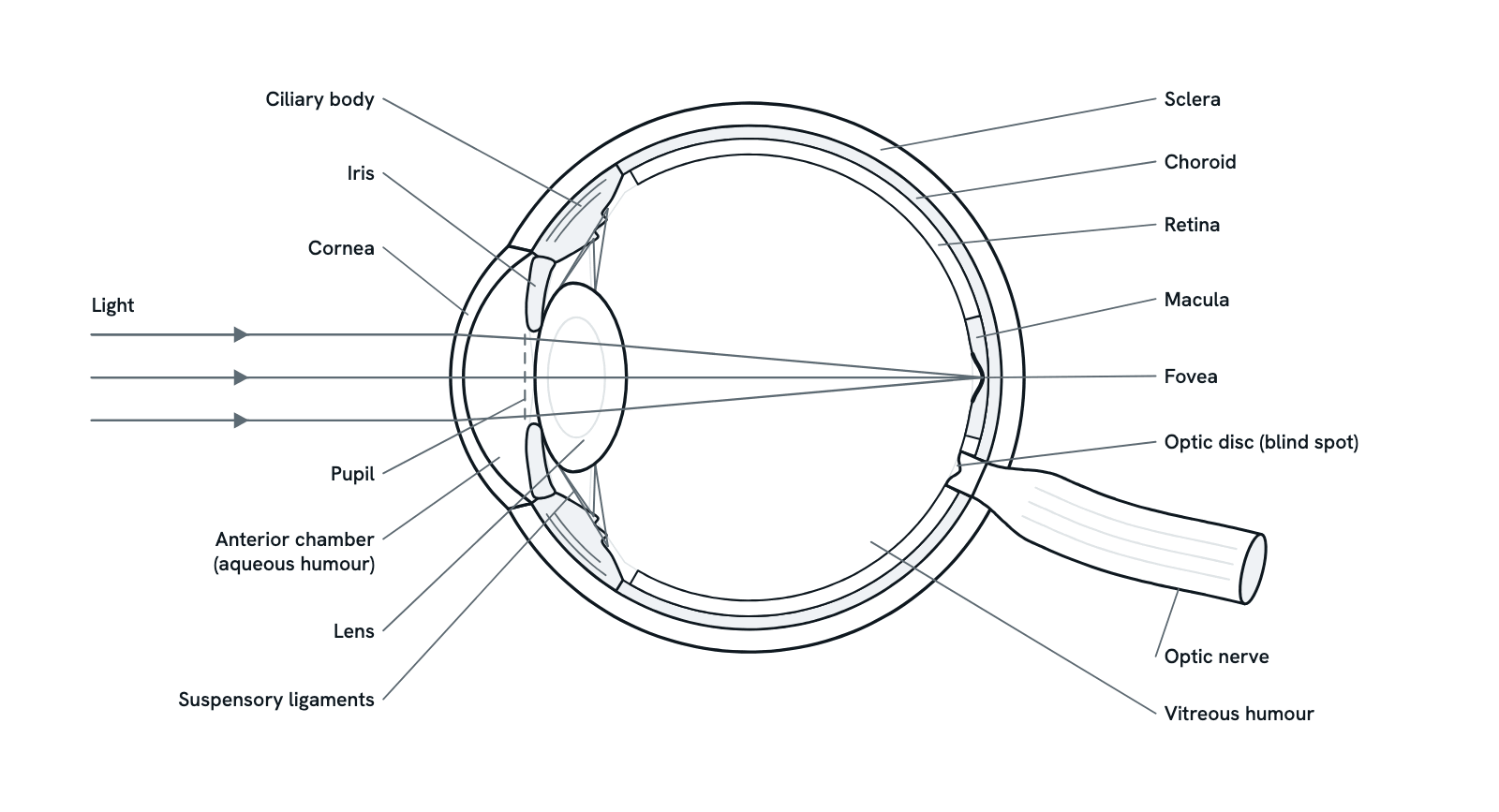
A horizontal section through the right eye, seen from above, with light entering from the left. The cornea and lens focus light on the retina, which turns it into nerve signals for the brain. Press any part to see what it is and what it does, or take the tour in order. Not to scale: the coats are drawn thicker than they are.
Also called Eye anatomy or Parts of the eye.
Press any part of the diagram, or pick one here, to see what it is and what it does.
Download the diagramChecked against its sources on 29 October 2026.
A tour
A tour of the human eye
01The eye at a glance
The eyeball is a fluid-filled sphere about 2.5 cm across. Its wall has three coats: the tough sclera outside, the choroid with its blood vessels in the middle, and the retina inside. At the front, the cornea, the pupil and the lens let light in and focus it on the retina.
02The cornea does most of the focusing
Light bends as it passes from air into the curved cornea, and this is where most of the eye's focusing happens. Under water this bending is almost lost, because water bends light about as much as the cornea does, which is why everything looks blurred there. The cornea's power is fixed.
03The iris sets the size of the pupil
Light passes through the aqueous humour of the anterior chamber and then the pupil, the opening in the iris. Muscles in the iris, under autonomic control, narrow the pupil in bright light and widen it in dim light. A smaller pupil also gives a sharper image.
04The lens fine-tunes the focus
Behind the pupil, the lens bends the light a little more, so that the rays from each point meet on the retina. The lens bends light much less than the cornea, but its power can change, so the eye can focus on objects at different distances.
05Accommodation: focusing near and far
For distant objects the ciliary muscle relaxes, the suspensory ligaments pull taut and the lens is held thin and flat. To focus on something near, the ring of ciliary muscle contracts, the ligaments slacken and the elastic lens rounds up, bending light more. This is accommodation.
06Two fluids keep the eye's shape
The ciliary body makes the watery aqueous humour, which flows through the pupil into the anterior chamber and drains at its angle. The balance between making and draining it keeps the pressure in the eye steady. Behind the lens, the vitreous humour, a clear jelly, fills about four fifths of the eye.
07The three coats
The sclera, the white of the eye, protects it and keeps its shape, and becomes the cornea at the front. The choroid's blood vessels feed the outer retina, and its pigment soaks up stray light. The retina, an outgrowth of the brain, is the only coat that senses light.
08Rods, cones and the fovea
In the retina, rods and cones turn light into electrical signals. There are about 91 million rods, which work in dim light, and about 4.5 million cones, which see colour and fine detail in bright light. The fovea, in the middle of the macula, is packed with cones, has no rods at its centre and gives the sharpest vision.
09The optic nerve and the blind spot
Light reaching the rods and cones changes how much transmitter they release onto the retina's other neurons. The ganglion cells then send action potentials along their axons, which leave the eye together at the optic disc as the optic nerve. The disc has no rods or cones, so it is the blind spot.
Parts
What each part of the human eye does
- Cornea
- The clear, dome-shaped front of the eye. Light bends most where it passes from air into the cornea, so the cornea does most of the eye's focusing, though its power is fixed.
- Aqueous humour
- A clear, watery fluid made by the ciliary body. It flows from the posterior chamber, behind the iris, through the pupil into the anterior chamber and drains at the angle where the iris meets the cornea. It feeds the cornea and lens.
- Iris
- The coloured ring of the eye, part of the middle coat. Two sets of muscle in it, with opposing actions and under autonomic control, narrow the pupil in bright light and widen it in dim light.
- Pupil
- The opening in the centre of the iris, through which light reaches the lens. A small pupil lets in less light but gives a sharper image with more depth of field.
- Lens
- A clear, elastic disc behind the pupil, held by the suspensory ligaments. It bends light less than the cornea does, but its power can change, which lets the eye focus on near and far objects.
- Ciliary body
- A ring of tissue round the lens, joining the iris to the choroid. Its ciliary muscle changes the shape of the lens, and its folds, the ciliary processes, make the aqueous humour.
- Suspensory ligaments
- Fine fibres, also called zonules, that run from the ciliary body to the edge of the lens. Pulled taut, they flatten the lens for distant vision; slackened, they let it round up for near vision.
- Vitreous humour
- A clear jelly that fills the space between the lens and the retina, about four fifths of the eye's volume. It helps keep the eye's shape and holds the retina against the choroid.
- Sclera
- The tough, white, fibrous outer coat: the white of the eye. It protects the inside of the eye, keeps its shape and gives the muscles that move the eye something to pull on. At the front it becomes the cornea.
- Choroid
- The middle coat at the back of the eye: a layer rich in blood vessels that feeds the outer retina, including the rods and cones. Its dark pigment absorbs stray light so it does not scatter inside the eye.
- Retina
- The inner coat, a thin sheet of nerve tissue that grows out from the brain. Its rods and cones turn light into electrical signals, which its other neurons process and pass to the ganglion cells, whose axons form the optic nerve.
- Macula
- A small area at the centre of the back of the retina, rich in cones, with the fovea at its middle. It gives the sharp, detailed central vision used for reading.
- Fovea
- A small pit at the centre of the macula, packed with cones, with no rods at all in its very centre, the foveola. The layers above the cones are pushed aside. Vision is sharpest here; we move our eyes to bring what we look at onto the fovea.
- Optic disc
- Where the axons of the retina gather and leave the eye as the optic nerve, and where the retina's main artery enters and its vein leaves. It has no rods or cones, so it cannot sense light: it is the blind spot.
- Optic nerve
- The bundle of axons of the retina's ganglion cells, carrying signals from the eye to the brain. The two optic nerves meet at the optic chiasm, and most signals then reach the visual cortex by way of the thalamus.
- Light rays
- Light from a distant point arrives in near-parallel rays. The cornea bends them most, the lens bends them a little more, and they meet on the retina at the fovea, forming a sharp image there.
Dictionary
The terms to know
Practise it
The quizzes and cards that fit
- Human Anatomy quizzes
- Shoulder and Rotator Cuff15 questions
- Knee Joint15 questions
- Arteries and Veins15 questions
- Neuroscience quizzes
- Reflex Arc15 questions
- Spinal Cord15 questions
- Cognitive Neuroscience: Language & Lateralization15 questions
- Physiology quizzes
- Exercise Physiology15 questions
- Puberty15 questions
- Blood Clotting15 questions
- Human Anatomy flashcards
The subject's terms as a ready-made deck, free to flip through.
Next to it
More diagrams
Sources
Where the facts come from
- National Eye Institute: How the Eyes Work
- StatPearls: Anatomy, Head and Neck, Eye (NCBI Bookshelf)
- Purves and others, Neuroscience, 2nd edition: Anatomy of the Eye (NCBI Bookshelf)
- Purves and others, Neuroscience, 2nd edition: The Formation of Images on the Retina (NCBI Bookshelf)
- Purves and others, Neuroscience, 2nd edition: Anatomical Distribution of Rods and Cones (NCBI Bookshelf)
- Purves and others, Neuroscience, 2nd edition: Central Projections of Retinal Ganglion Cells (NCBI Bookshelf)
- Hussey and others, Patterning and Development of Photoreceptors in the Human Retina, Front Cell Dev Biol 2022 (PMC)
Drawn and written by biofideX from these sources.