Human Ear Diagram
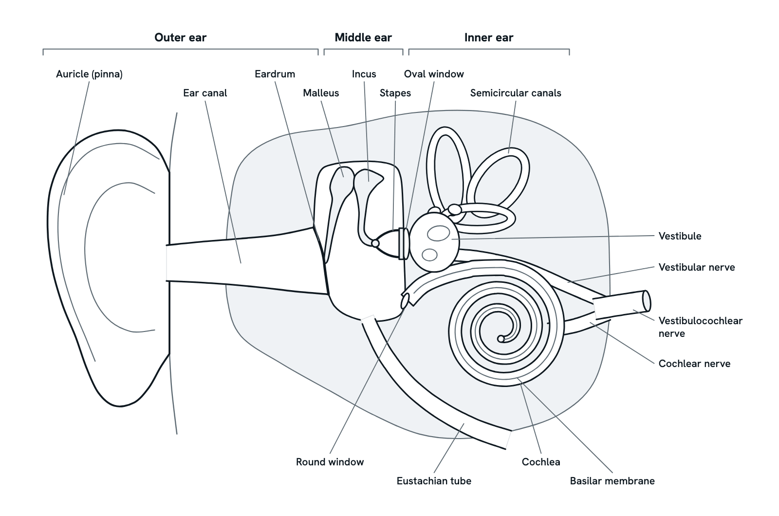
The parts of the human ear and what each does: the auricle, ear canal, eardrum, ossicles, cochlea, semicircular canals and the nerve to the brain.
A coronal section through the right ear, seen from the front, with the outer ear on the left and the inner ear on the right. Sound passes inward through all three parts, and the inner ear also senses balance. Press any part to see what it is and what it does, or take the tour in order. Not to scale: the middle and inner ear are drawn larger.
Also called Ear anatomy or Parts of the ear.
Press any part of the diagram, or pick one here, to see what it is and what it does.
Download the diagramChecked against its sources on 29 October 2026.
A tour
A tour of the human ear
01The ear at a glance
The ear has three parts. The outer ear collects sound; the air-filled middle ear carries it inward on three tiny bones; and the fluid-filled inner ear, deep in the temporal bone, turns it into nerve signals. The inner ear also holds the organs of balance.
02Sound is collected
Sound waves are vibrations passing through the air. The auricle gathers them and funnels them into the ear canal, a narrow passage that carries them to the eardrum.
03The eardrum vibrates
The sound waves make the eardrum vibrate: faster for high-pitched sounds, slower for low ones, and further for loud ones. The handle of the malleus is attached to the eardrum, so the vibrations pass straight to the first of the three ossicles.
04The ossicles boost the pressure
In the air-filled middle ear, the malleus moves the incus, and the incus moves the stapes, whose footplate sits in the oval window. The force gathered by the large eardrum is passed, with some lever action from the ossicles, onto the far smaller footplate, so the pressure rises. This helps the vibrations pass from the air into the fluid of the inner ear.
05The Eustachian tube balances the pressure
The Eustachian tube runs from the middle ear to the upper part of the throat. It lets air in or out, so the pressure behind the eardrum stays level with the air outside, and it lets fluid drain from the middle ear. When it is blocked, as in a cold, fluid can build up there.
06A wave runs through the cochlea
As the stapes rocks in the oval window, it makes the fluid inside the cochlea ripple, and a travelling wave runs along the basilar membrane. Each time the stapes pushes in, the membrane of the round window bulges out, giving the fluid room to move.
07Hair cells turn movement into signals
Hair cells sit on the basilar membrane and ride the wave. As the tiny hair-like stereocilia on top of each cell bend, channels at their tips open, ions rush in and an electrical signal is made. High pitches move the membrane most near the base of the cochlea, where it is narrow and stiff; low pitches move it most near the apex, where it is wide and floppy.
08Signals travel to the brain
The hair cells release a neurotransmitter onto fibres of the cochlear nerve, which fire action potentials. The cochlear nerve joins the vestibular nerve as the vestibulocochlear nerve, the eighth cranial nerve, which carries the signals to the brainstem. From there they are relayed up to the auditory cortex, where they are recognised as sound.
09Balance: the vestibule and semicircular canals
Hair cells also sense the head's position and movement. In the three semicircular canals, moving fluid bends them when the head turns. In the utricle and saccule of the vestibule, tiny crystals pulled by gravity bend them when the head tilts or moves in a straight line. The vestibular nerve carries these signals to the brain.
Parts
What each part of the human ear does
- Auricle (pinna)
- The visible outer ear: a curved flap of cartilage covered with skin. It gathers sound waves and funnels them into the ear canal.
- Ear canal
- A narrow passage that carries sound from the auricle to the eardrum. Glands in the skin lining its outer part make earwax (cerumen), which helps keep it clean.
- Eardrum (tympanic membrane)
- A thin membrane across the inner end of the ear canal, between the outer and middle ear. Sound waves make it vibrate, and the handle of the malleus, attached to it, picks up the vibrations.
- Malleus (hammer)
- The first of the three ossicles, the tiny bones of the middle ear. Its handle is attached to the eardrum and its head joins the incus, so it moves with every vibration of the eardrum.
- Incus (anvil)
- The middle ossicle. Its body joins the head of the malleus and its long process joins the stapes, passing the vibrations from one to the other.
- Stapes (stirrup)
- The last ossicle and the smallest bone in the body. Its footplate sits in the oval window, so as it rocks it pushes the fluid of the inner ear in and out.
- Eustachian tube
- A narrow passage, also called the auditory tube, from the middle ear to the upper part of the throat. It lets air in and out, keeping the pressure on both sides of the eardrum equal, and lets fluid drain from the middle ear.
- Oval window
- An opening from the middle ear into the vestibule of the inner ear, filled by the footplate of the stapes. As the stapes moves, the oval window passes its vibrations to the fluid inside the cochlea.
- Round window
- A membrane-covered opening below the oval window, at the base of the cochlea's lower channel. The fluid in the cochlea cannot be squeezed, so the round window bulges out as the stapes pushes in, giving the fluid room to move.
- Vestibule
- The central chamber of the inner ear, just inside the oval window. Its two otolith organs, the utricle and saccule, sense the pull of gravity and movement of the head in a straight line.
- Semicircular canals
- Three fluid-filled loops set at roughly right angles to one another. When the head turns, the fluid in them moves and bends a jelly-like cap over hair cells in the swelling (ampulla) at the base of each canal.
- Cochlea
- The snail-shaped, fluid-filled organ of hearing. Vibrations from the stapes make its fluid ripple, setting up a travelling wave along the basilar membrane inside it.
- Basilar membrane
- An elastic partition that runs the length of the cochlea and divides it into upper and lower parts. The hair cells sit on it: those near the base of the cochlea, where the membrane is narrow and stiff, respond to high pitches; those near the apex, where it is wide and floppy, to low ones.
- Vestibular nerve
- The branch of the vestibulocochlear nerve that carries signals about balance and movement of the head from the vestibule and the semicircular canals.
- Cochlear nerve
- The branch of the vestibulocochlear nerve that carries signals about sound from the hair cells of the cochlea. It is also called the auditory nerve.
- Vestibulocochlear nerve
- The eighth cranial nerve, formed as the cochlear and vestibular nerves run together. It leaves the inner ear through a canal in the temporal bone and enters the brainstem, where its fibres reach the cochlear and vestibular nuclei.
Dictionary
The terms to know
Practise it
The quizzes and cards that fit
- Human Anatomy quizzes
- Shoulder and Rotator Cuff15 questions
- Knee Joint15 questions
- Arteries and Veins15 questions
- Neuroscience quizzes
- Reflex Arc15 questions
- Spinal Cord15 questions
- Cognitive Neuroscience: Language & Lateralization15 questions
- Physiology quizzes
- Exercise Physiology15 questions
- Puberty15 questions
- Blood Clotting15 questions
- Human Anatomy flashcards
The subject's terms as a ready-made deck, free to flip through.
Next to it
More diagrams
Sources
Where the facts come from
- National Institute on Deafness and Other Communication Disorders: How Do We Hear?
- National Institute on Deafness and Other Communication Disorders: Balance Disorders
- National Institute on Deafness and Other Communication Disorders: Ear Infections in Children
- StatPearls: Neuroanatomy, Ear (NCBI Bookshelf)
- StatPearls: Anatomy, Head and Neck, Ear Ossicles (NCBI Bookshelf)
- StatPearls: Physiology, Ear (NCBI Bookshelf)
- StatPearls: Middle Ear Anatomy and Physiology (NCBI Bookshelf)
- StatPearls: Inner Ear Anatomy and Cochlear Physiology (NCBI Bookshelf)
- Ugarteburu and others, Mammalian Middle Ear Mechanics: A Review, Front Bioeng Biotechnol 2022 (PMC)
- Wolf and others, Is there an unmet medical need for improved hearing restoration? EMBO Mol Med 2022 (PMC)
Drawn and written by biofideX from these sources.