Skin Diagram
The layers of the skin and what each does: epidermis, dermis and hypodermis, with hair, glands, blood vessels, nerves and touch receptors.
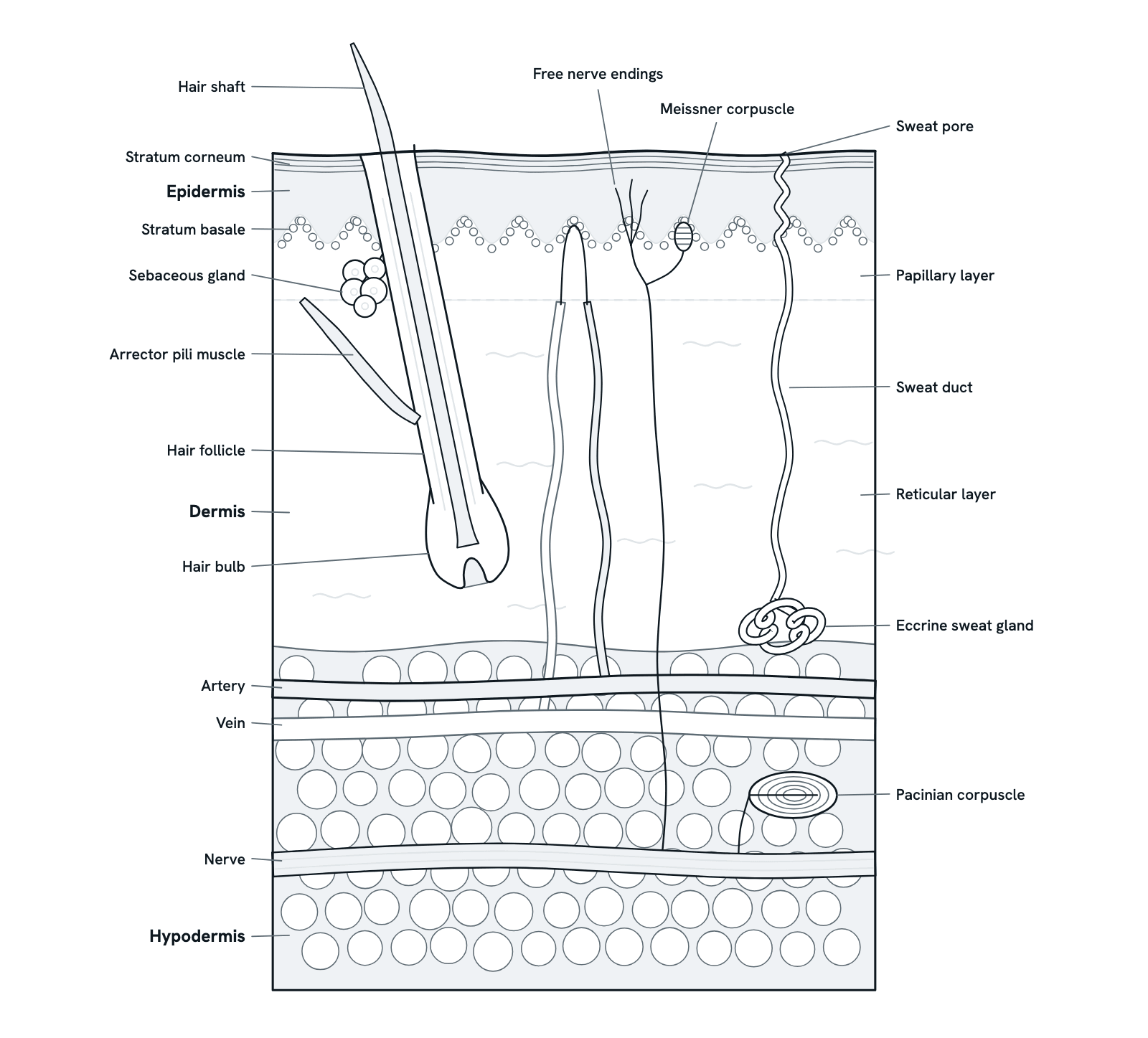
Skin has three layers: the epidermis on top, the dermis beneath it and the fatty hypodermis below. Between them they protect the body, hold its temperature steady and sense touch, pressure, heat and pain. Press any part, or take the tour. A block of thin, hairy skin in section, not to scale.
Also called Layers of the skin, Skin anatomy or Integumentary system.
Press any part of the diagram, or pick one here, to see what it is and what it does.
Download the diagramChecked against its sources on 29 October 2026.
A tour
A tour of the skin
01Skin at a glance
Skin is the body's largest organ. It has three layers: the epidermis, an outer sheet of cells; the dermis, a tough layer of connective tissue with vessels, nerves, glands and hair follicles; and the hypodermis of fat beneath. It protects, keeps temperature steady, senses the outside world and makes vitamin D.
02The epidermis
Stem cells in the stratum basale divide, and their daughters move up through the stratum spinosum and stratum granulosum, flattening and filling with keratin. By the time they reach the stratum corneum they are dead, flat and tough, and they are shed from the surface. Thick skin on the palms and soles has one more layer, the stratum lucidum, just under the corneum. The epidermis has no blood vessels; it is fed from the dermis below.
03The dermis
The dermis is connective tissue rich in collagen and elastic fibres. Its thin papillary layer rises into the epidermis as dermal papillae, holding small blood vessels and touch receptors. The thicker reticular layer gives skin its strength and holds the hair follicles, glands, vessels and nerves.
04Protection
The stratum corneum and its lipids form a barrier that keeps water in and microbes, chemicals and wear out. Melanin from melanocytes in the stratum basale shields the cells beneath from ultraviolet light, and sebum from sebaceous glands keeps the surface supple and helps keep microbes in check.
05Temperature control
When the body is hot, eccrine glands release sweat, which cools the skin as it evaporates, and vessels in the dermis widen so more warm blood flows near the surface and loses heat. In the cold, the vessels narrow to keep heat in, and arrector pili muscles raise the hairs as goosebumps.
06Sensation
Sensory endings at different depths answer to different things. Free nerve endings in the epidermis sense pain, heat and cold. Meissner corpuscles in the dermal papillae, most of all in hairless skin such as the fingertips, sense light touch, and Pacinian corpuscles deep in the skin sense pressure and vibration. Their fibres run back in nerves to the spinal cord.
07Hair and its glands
Each hair grows from the bulb at the base of its follicle, where dividing cells make new hair from below. A sebaceous gland empties into the follicle, and an arrector pili muscle runs from the follicle to the upper dermis. Thin skin over most of the body has hair; the palms and soles have none.
08The hypodermis
Below the dermis, the hypodermis (subcutaneous tissue) is mostly fat held in connective tissue. It insulates the body and keeps heat in, cushions the organs beneath against knocks, stores energy as fat, and carries the larger vessels and nerves that supply the skin.
Parts
What each part of the skin does
- Epidermis
- The outer layer: a stratified epithelium made mostly of keratinocytes. It has no blood vessels of its own and is fed from the dermis below. It is thickest on the palms and soles.
- Stratum corneum
- The surface layer of the epidermis: many layers of flat, dead keratinocytes filled with keratin and sealed with lipids. It keeps water in and microbes out, and is shed and replaced all the time.
- Stratum basale
- The deepest layer of the epidermis, resting on the basement membrane. Its stem cells divide to make new keratinocytes, which move up and mature; melanocytes here make the pigment melanin.
- Papillary layer of the dermis
- The thin upper layer of the dermis, of loose connective tissue. It rises into the epidermis as dermal papillae, which hold small blood vessels that feed the epidermis, and touch receptors.
- Reticular layer of the dermis
- The thick lower layer of the dermis: dense connective tissue of collagen bundles and elastic fibres that gives skin its strength and stretch. It holds hair follicles, glands, blood vessels and nerves.
- Hypodermis
- The subcutaneous layer below the dermis, mostly fat cells (adipose tissue) with connective tissue. It insulates the body, cushions it against knocks and stores energy as fat.
- Hair
- A thread made largely of the protein keratin. The shaft is the part above the skin; below the surface the hair continues as its root, inside the follicle.
- Hair follicle
- A tube of epithelium that grows down from the epidermis into the dermis and holds the root of the hair. Its upper part opens at the skin surface, where sebum from its gland comes out.
- Hair bulb
- The swollen base of the follicle, where dividing matrix cells make the hair. A dermal papilla of connective tissue and capillaries pushes up into it from below and supports its growth.
- Sebaceous gland
- An oil gland that empties into the hair follicle. Its cells fill with sebum, a fatty secretion, and break down to release it; sebum keeps skin and hair supple and helps keep microbes in check.
- Arrector pili muscle
- A small band of smooth muscle from the follicle to the upper dermis. When it contracts, in the cold for example, it pulls the hair upright and raises a goosebump; it also squeezes the sebaceous gland.
- Eccrine sweat gland
- A coiled tube deep in the dermis or upper hypodermis that makes watery sweat. Sympathetic nerves set it working when the body is hot, and sweat cools the skin as it evaporates.
- Sweat duct and pore
- The duct carries sweat from the coiled gland up through the dermis and on through the epidermis, to open as a pore on the surface.
- Meissner corpuscle
- A small capsule of stacked cells round nerve endings, sitting in a dermal papilla just under the epidermis. It senses light touch and gentle, low-frequency vibration. Meissner corpuscles belong mainly to hairless skin, such as the fingertips, palms, soles and lips.
- Pacinian corpuscle
- Also called a lamellated corpuscle: a large, onion-like capsule of many layers round a nerve ending, deep in the skin. It senses deep pressure and vibration.
- Free nerve endings
- Bare branches of sensory nerve fibres that reach up into the epidermis. They sense pain, heat and cold, and some sense touch.
- Nerve
- A bundle of nerve fibres (axons) in the deep skin. Sensory fibres carry signals from the skin's receptors to the spinal cord; sympathetic fibres run out to blood vessels, sweat glands and arrector pili muscles.
- Artery and capillaries
- An artery in the hypodermis sends branches up through the dermis to capillary loops in the dermal papillae, which feed the epidermis. Widening or narrowing these vessels lets the skin lose or keep heat.
- Vein
- Collects blood from the capillaries of the dermis through small veins (venules) and carries it away. Veins and arteries run together in networks at the base of the dermis and beneath the papillae.
Dictionary
The terms to know
Practise it
The quizzes and cards that fit
- Human Anatomy quizzes
- Shoulder and Rotator Cuff15 questions
- Knee Joint15 questions
- Arteries and Veins15 questions
- Physiology quizzes
- Exercise Physiology15 questions
- Puberty15 questions
- Blood Clotting15 questions
- Human Anatomy flashcards
The subject's terms as a ready-made deck, free to flip through.
Next to it
More diagrams
Sources
Where the facts come from
- StatPearls: Anatomy, Skin (Integument) (NCBI Bookshelf)
- StatPearls: Anatomy, Skin (Integument), Epidermis (NCBI Bookshelf)
- StatPearls: Histology, Skin Appendages (NCBI Bookshelf)
- StatPearls: Anatomy, Skin, Sweat Glands (NCBI Bookshelf)
- StatPearls: Histology, Meissner Corpuscle (NCBI Bookshelf)
- NCI SEER Training: Anatomy of the Skin
- NCI SEER Training: Layers of the Skin
Drawn and written by biofideX from these sources.