Bones of the Foot Diagram
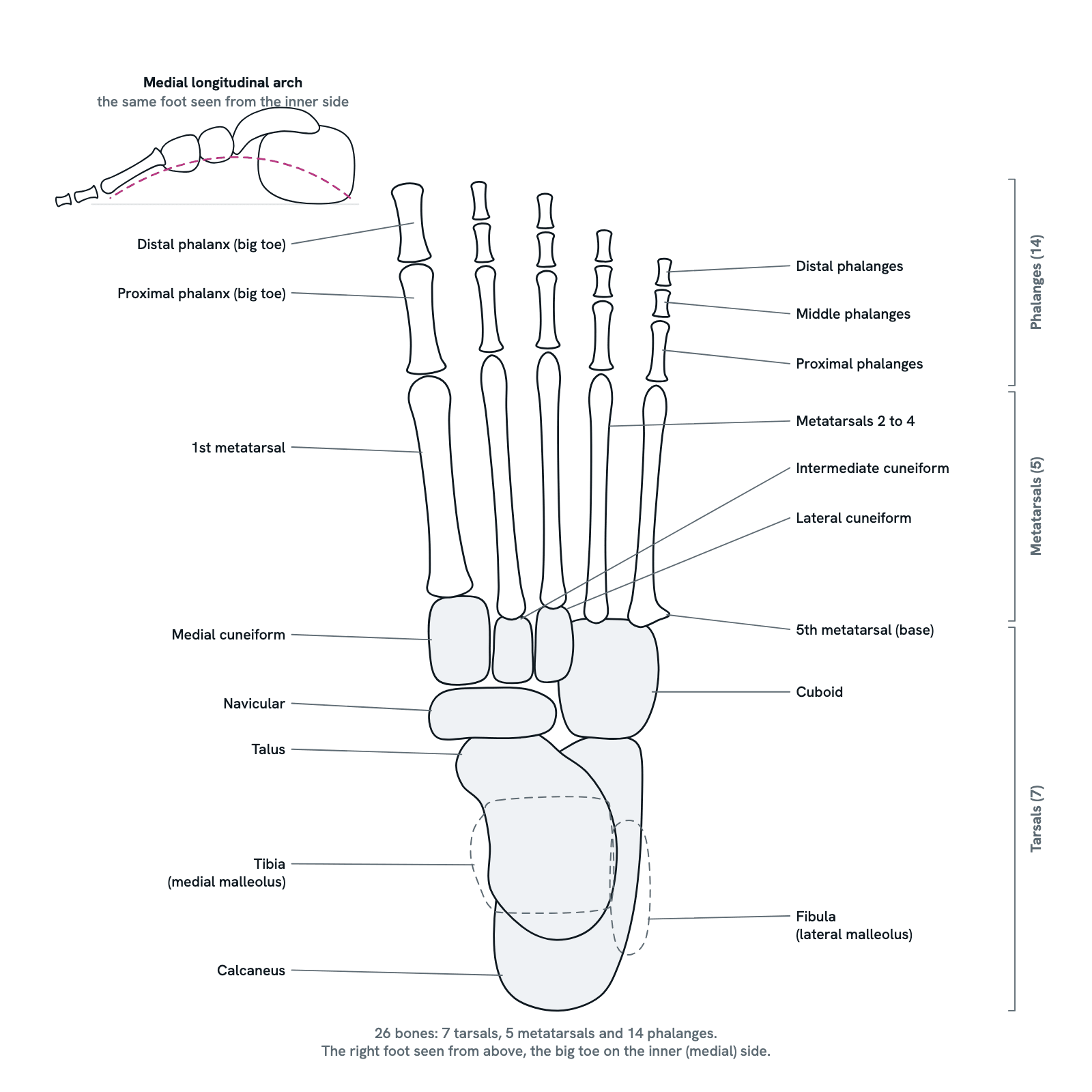
The 26 bones of the foot labelled: 7 tarsals, 5 metatarsals and 14 phalanges, with the joints between them, the arches and the common fractures.
The right foot seen from above, toes at the top and the big toe on the inner side. Seven tarsal bones make the heel and the midfoot, five metatarsals the sole, and fourteen phalanges the toes: 26 bones in all. The ends of the tibia and fibula are drawn faintly where they grip the talus at the ankle. Press any bone to see what it joins and what goes wrong with it, or take the tour.
Also called Foot bones, Foot anatomy, Tarsal bones or Foot skeleton.
Press any part of the diagram, or pick one here, to see what it is and what it does.
Download the diagramChecked against its sources on 5 November 2026.
A tour
A tour of the bones of the foot
0126 bones in three regions
The foot has 26 bones in three regions. The hindfoot is the talus and the calcaneus; the midfoot is the navicular, the cuboid and the three cuneiforms; together these seven are the tarsals. The forefoot is the five metatarsals and the fourteen phalanges of the toes. Between them are some 30 joints, each surface covered in cartilage, which is what lets a rigid lever also be a spring.
02The hindfoot: talus, calcaneus and two joints
The talus is the only foot bone that meets the leg: its dome fits into the mortise between the malleoli of the tibia and fibula, a hinge for pointing the foot up (dorsiflexion) and down (plantarflexion). Beneath it the calcaneus carries the body's weight to the ground and forms the heel. The subtalar joint between the two turns the sole in and out: inversion and eversion.
03The midfoot and the transverse tarsal joint
In front of the hindfoot, the navicular takes the head of the talus on the inner side and the cuboid continues from the calcaneus on the outer side. These two joints together, talonavicular and calcaneocuboid, form the transverse tarsal (Chopart) joint, which adds to inversion and eversion. The three cuneiforms, wedges set side by side in front of the navicular, complete the midfoot.
04The forefoot: metatarsals and phalanges
Five metatarsals, numbered 1 to 5 from the big toe, run from the midfoot to the toes, each with a base, a shaft and a rounded head. Each toe then has a proximal, a middle and a distal phalanx, except the big toe (hallux), which has only a proximal and a distal one: 14 phalanges, and two joints in the big toe where the other toes have three.
05The Lisfranc joint, where the midfoot meets the metatarsals
The bases of the five metatarsals meet the three cuneiforms and the cuboid at the tarsometatarsal, or Lisfranc, joint. The base of the 2nd metatarsal sits in a slot between the medial and lateral cuneiforms, tied to the medial cuneiform by the Lisfranc ligament: the keystone of the midfoot. An axial load on a foot pointed down, as in a tackle or a car crash, can sprain or break it. Bruising on the sole is the warning sign, and about 20% are missed at first.
06The arches of the foot
The bones are set in arches, not flat. The medial longitudinal arch, the highest, runs from the heel through the talus, navicular and cuneiforms to the inner metatarsals; the lateral arch from the heel through the cuboid to the outer two; a transverse arch crosses the midfoot. The plantar fascia and ligaments beneath and tendons such as tibialis posterior hold them up. Where the medial arch sinks the foot is flat, pes planus: normal in a baby, often harmless in adults.
07Walking: heel strike to toe-off
Each step begins with the heel striking the ground, so the calcaneus takes the first load. In midstance the whole foot is down, the arch sinks a little and the plantar fascia stretches and stores energy. Then the heel rises, the toes bend up at the metatarsophalangeal joints and the fascia, fixed to the proximal phalanges, winds tight and raises the arch, the windlass mechanism, so the foot becomes a stiff lever for the push-off.
08A mnemonic for the seven tarsals
Tiger Cubs Need MILC: Talus, Calcaneus, Navicular, then the Medial, Intermediate and Lateral cuneiforms, then the Cuboid. It names the hindfoot first, then the midfoot from the inner side outwards. Another way to place them runs from the heel forwards: the talus sits on the calcaneus, the navicular is in front of the talus, the cuboid in front of the calcaneus, and the three cuneiforms in front of the navicular.
09The big toe: sesamoids and bunions
Under the head of the 1st metatarsal lie two sesamoid bones, embedded in the tendons beneath the joint as pulleys and seen from the sole rather than from above. At the first metatarsophalangeal joint a bunion, hallux valgus, is the big toe drifting outwards while the metatarsal drifts inwards, so its head stands out on the inner side. It affects about a quarter of adults, women far more than men; wider shoes come first and surgery is for pain that persists, not for looks.
10Common injuries, and which are bone
A sprained ankle is a torn ligament, not a broken bone: most often the anterior talofibular ligament, when the foot rolls inwards. The same inversion can pull the tuberosity off the base of the 5th metatarsal, the most often fractured metatarsal. Repeated loading cracks the 2nd metatarsal, the usual site of a metatarsal stress fracture. A fall onto the heel breaks the calcaneus through the subtalar joint, and a displaced fracture of the talar neck can cut off its blood.
Parts
What each part of the bones of the foot does
- Talus
- The ankle bone. Its dome sits in the mortise of the tibia and fibula, its body rests on the calcaneus at the subtalar joint and its head meets the navicular. About 60% of its surface is cartilage and no muscle or tendon attaches to it, so its blood supply is fragile: a displaced neck fracture risks avascular necrosis.
- Calcaneus
- The heel bone, the largest tarsal. It carries the talus on three facets, meets the cuboid in front, takes the Achilles tendon at its tuberosity and gives origin to the plantar fascia. A fall onto the heel drives the talus down into it; about three quarters of its fractures run into the subtalar joint.
- Navicular
- On the inner side of the midfoot, between the head of the talus and the three cuneiforms, with the cuboid beside it. The tibialis posterior tendon, which helps hold up the medial longitudinal arch, inserts on its tuberosity. Its blood supply is poor, so its stress fractures heal slowly and may not unite.
- Cuboid
- The cube-shaped bone of the outer midfoot, between the calcaneus behind and the bases of the 4th and 5th metatarsals in front, with the navicular and the lateral cuneiform on its inner side. A groove on its underside carries the peroneus longus tendon across the sole to the inner side of the foot.
- Medial cuneiform
- The innermost of the three wedge-shaped cuneiforms, between the navicular and the base of the 1st metatarsal. The tibialis anterior tendon inserts on it, and the Lisfranc ligament runs from it to the base of the 2nd metatarsal, the strongest tie holding the midfoot together.
- Intermediate cuneiform
- The middle wedge, between the navicular and the 2nd metatarsal. Its front end lies behind those of its neighbours, so the base of the 2nd metatarsal sits in a slot between the medial and lateral cuneiforms: the keystone that locks the Lisfranc joint.
- Lateral cuneiform
- The outer wedge, between the navicular behind and the 3rd metatarsal in front, with the intermediate cuneiform on its inner side and the cuboid on its outer side. The three cuneiforms and the cuboid form the row the metatarsals stand on, the tarsometatarsal or Lisfranc joint.
- 1st metatarsal
- The thick metatarsal of the big toe, from the medial cuneiform to the proximal phalanx. Two sesamoid bones lie under its head, which this view from above hides. In hallux valgus the metatarsal drifts inwards and the big toe outwards, and the head stands out on the inner side of the foot as a bunion.
- 2nd, 3rd and 4th metatarsals
- Long bones, each with a base on the midfoot (the 2nd and 3rd on the cuneiforms, the 4th on the cuboid), a slender shaft and a rounded head that meets the proximal phalanx. The 2nd is set deepest into the midfoot and is the commonest site of a stress fracture, the march fracture of soldiers and runners.
- 5th metatarsal
- The outer metatarsal, with a tuberosity at its base where the peroneus brevis tendon inserts. It is the metatarsal most often broken: an inversion injury pulls the tuberosity off, and a break at the junction of base and shaft (a Jones fracture) heals poorly, as the blood supply there is a watershed.
- Proximal phalanges
- The first bone of each toe, meeting the head of its metatarsal at the metatarsophalangeal (MTP) joint. The plantar fascia runs from the heel to their bases, so when the toes are bent up at the end of a step it tightens and pulls the arch higher: the windlass mechanism.
- Middle phalanges
- The second bone of toes 2 to 5. The big toe has none: it has two phalanges and two joints where the other toes have three, which is why there are 14 phalanges and not 15.
- Distal phalanges
- The last bone of each toe, under the nail. The big toe's is the broadest; those of the other toes are small, and the little toe's smallest of all.
- Ankle: tibia and fibula
- Drawn faintly: the ends of the two leg bones grip the talus between their malleoli to form the ankle joint, a hinge that points the foot up (dorsiflexion) and down (plantarflexion). Turning the sole in and out, inversion and eversion, happens lower down, at the subtalar joint between talus and calcaneus.
- Medial longitudinal arch
- Seen from the inner side: calcaneus, talus, navicular, cuneiforms and the inner metatarsals form a curve that rests on the ground only at the heel and the first three metatarsal heads. The plantar fascia, the spring ligament and the tibialis posterior tendon hold it up; when it sinks the foot is flat (pes planus).
- Three regions, 26 bones
- Seven tarsals make the hindfoot (talus and calcaneus) and the midfoot (navicular, cuboid and three cuneiforms); five metatarsals and fourteen phalanges make the forefoot. The two sesamoids under the head of the 1st metatarsal are extra, and not counted.
As a table
The bones of the foot as a table
The bones of the foot by region, what each articulates with, and a point worth knowing about each.
Talus
- Region
- Hindfoot (tarsal)
- Articulates with
- Tibia and fibula (ankle), calcaneus (subtalar), navicular
- Note
- No muscle attaches to it; about 60% is cartilage; fractures risk avascular necrosis
Calcaneus
- Region
- Hindfoot (tarsal)
- Articulates with
- Talus (three facets), cuboid
- Note
- Largest tarsal; Achilles tendon and plantar fascia attach to it; broken by falls onto the heel
Navicular
- Region
- Midfoot (tarsal)
- Articulates with
- Talus, three cuneiforms, cuboid
- Note
- Tibialis posterior inserts on it; poor blood supply, so stress fractures heal slowly
Cuboid
- Region
- Midfoot (tarsal)
- Articulates with
- Calcaneus, 4th and 5th metatarsals, lateral cuneiform, navicular
- Note
- Grooved underneath for the peroneus longus tendon
Medial cuneiform
- Region
- Midfoot (tarsal)
- Articulates with
- Navicular, 1st and 2nd metatarsals, intermediate cuneiform
- Note
- Tibialis anterior inserts on it; the Lisfranc ligament runs from it to the 2nd metatarsal
Intermediate cuneiform
- Region
- Midfoot (tarsal)
- Articulates with
- Navicular, 2nd metatarsal, the other cuneiforms
- Note
- Set back, so the 2nd metatarsal base sits between its neighbours: the keystone of the Lisfranc joint
Lateral cuneiform
- Region
- Midfoot (tarsal)
- Articulates with
- Navicular, 2nd and 3rd metatarsals, intermediate cuneiform, cuboid
- Note
- Outer wedge of the three
1st metatarsal
- Region
- Forefoot
- Articulates with
- Medial cuneiform, proximal phalanx of the big toe
- Note
- Two sesamoids under its head; hallux valgus (bunion) at its joint with the big toe
2nd to 4th metatarsals
- Region
- Forefoot
- Articulates with
- Cuneiforms (2nd, 3rd) or cuboid (4th), and the proximal phalanges
- Note
- The 2nd is the commonest site of a stress (march) fracture
5th metatarsal
- Region
- Forefoot
- Articulates with
- Cuboid, proximal phalanx of the little toe
- Note
- Most often fractured; peroneus brevis inserts on its tuberosity; Jones fracture at the base-shaft junction
Proximal phalanges (5)
- Region
- Forefoot
- Articulates with
- Metatarsal heads (MTP joints), middle or distal phalanges
- Note
- The plantar fascia attaches to their bases: the windlass mechanism
Middle phalanges (4)
- Region
- Forefoot
- Articulates with
- Proximal and distal phalanges of toes 2 to 5
- Note
- The big toe has none
Distal phalanges (5)
- Region
- Forefoot
- Articulates with
- Middle phalanges, or the proximal phalanx in the big toe
- Note
- Carry the nails; the big toe's is broad
Sesamoids (2)
- Region
- Forefoot, extra
- Articulates with
- Under the head of the 1st metatarsal
- Note
- Embedded in tendon, act as pulleys; not counted among the 26
The seven tarsals, five metatarsals and fourteen phalanges make 26 bones. Counts of joints vary with how they are counted, from about 30 to 33.
Dictionary
The terms to know
Practise it
The quizzes and cards that fit
- Human Anatomy quizzes
- Shoulder and Rotator Cuff15 questions
- Knee Joint15 questions
- Arteries and Veins15 questions
- Medicine quizzes
- Labor and Delivery15 questions
- IV Fluids15 questions
- Sepsis15 questions
- Human Anatomy flashcards
The subject's terms as a ready-made deck, free to flip through.
Next to it
More diagrams
Sources
Where the facts come from
- StatPearls: Anatomy, Bony Pelvis and Lower Limb: Foot and Ankle (NCBI Bookshelf)
- StatPearls: Anatomy, Appendicular Skeleton (NCBI Bookshelf)
- StatPearls: Anatomy, Bony Pelvis and Lower Limb, Foot Joints (NCBI Bookshelf)
- StatPearls: Anatomy, Bony Pelvis and Lower Limb: Arches of the Foot (NCBI Bookshelf)
- StatPearls: Anatomy, Bony Pelvis and Lower Limb: Navicular Bone (NCBI Bookshelf)
- StatPearls: Anatomy, Bony Pelvis and Lower Limb: Foot Cuboid Bone (NCBI Bookshelf)
- StatPearls: Anatomy, Sesamoid Bones (NCBI Bookshelf)
- StatPearls: Calcaneus Fractures (NCBI Bookshelf)
- StatPearls: Talus Fracture (NCBI Bookshelf)
- StatPearls: Tarsal Navicular Fractures (NCBI Bookshelf)
- StatPearls: Lisfranc Dislocation (NCBI Bookshelf)
- StatPearls: 5th Metatarsal Fracture (NCBI Bookshelf)
- StatPearls: March Fracture (Metatarsal Stress Fractures) (NCBI Bookshelf)
- StatPearls: Hallux Valgus (NCBI Bookshelf)
- StatPearls: Pes Planus (NCBI Bookshelf)
- StatPearls: Acute Ankle Sprain (NCBI Bookshelf)
- The Diagnosis, Management and Complications Associated with Fractures of the Talus, Open Orthopaedics Journal 2017 (PMC)
- Intra-articular Calcaneus Fractures: Current Concepts Review, Foot and Ankle Orthopaedics 2020 (PMC)
- Lisfranc fracture-dislocations: current management, EFORT Open Reviews 2019 (PMC)
- Lisfranc Injury: Recent Trends in Management, Cureus 2023 (PMC)
- Fifth metatarsal fractures: an update on management, complications, and outcomes, EFORT Open Reviews 2022 (PMC)
- Hallux Valgus, Foot and Ankle Orthopaedics 2019 (PMC)
- Acute ankle sprain in athletes: Clinical aspects and algorithmic approach, World Journal of Orthopedics 2020 (PMC)
- Stress Fractures of the Foot and Ankle in Athletes, Foot and Ankle Orthopaedics 2023 (PMC)
- The rise of the longitudinal arch when sitting, standing, and walking: Contributions of the windlass mechanism, PLoS One 2021 (PMC)
- Evolutionary anatomy of the plantar aponeurosis in primates, including humans, Journal of Anatomy 2020 (PMC)
- Tibialis posterior in health and disease: a review of structure and function, Journal of Foot and Ankle Research 2009 (PMC)
- Anatomical Description of the Spring Ligament Articular Facet, Foot and Ankle Orthopaedics 2024 (PMC)
- Imaging Anatomy of the Ankle in Normal and Pathological States, Cureus 2025 (PMC)
Drawn and written by biofideX from these sources.