Carpal Bones Diagram
The eight carpal bones of the wrist in two rows, with the radius, ulna and metacarpals, the mnemonic and the carpal tunnel.
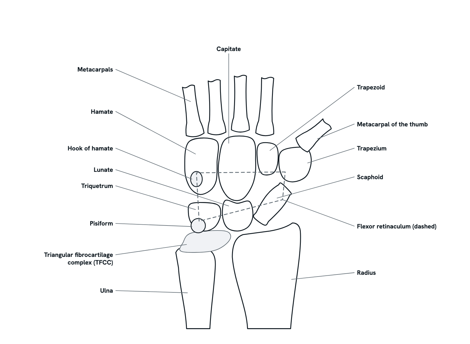
The right wrist seen from the palm, thumb to the right: the radius and ulna below, the eight carpal bones in two rows of four, and the bases of the five metacarpals above. The dashed band is the flexor retinaculum, the roof of the carpal tunnel. Press any bone for its shape, its neighbours and its clinical point, or take the tour, which starts with the mnemonic. Not to scale.
Also called Wrist bones, Bones of the wrist or Carpal bones mnemonic.
Press any part of the diagram, or pick one here, to see what it is and what it does.
Download the diagramChecked against its sources on 5 November 2026.
A tour
A tour of the carpal bones
01The wrist at a glance
Eight carpal bones link the forearm to the hand, between the radius and ulna below and the bases of the five metacarpals above. They lie in two rows of four: a proximal row that meets the radius, and a distal row that is bound to the metacarpals. This is the right wrist seen from the palm, so the thumb is on the right.
02Naming the eight: Some Lovers Try Positions That They Cannot Handle
Read from the thumb side to the little finger, the proximal row is Scaphoid, Lunate, Triquetrum, Pisiform, and the distal row is Trapezium, Trapezoid, Capitate, Hamate. The mnemonic gives their first letters in that order: Some Lovers Try Positions That They Cannot Handle. In the drawing it runs right to left along each row, because the thumb is on the right.
03The radiocarpal joint: the radius, not the ulna
The wrist joint is the radiocarpal joint, a condyloid synovial joint. The scaphoid and lunate rest on the distal end of the radius. The ulna does not reach the carpals: between its head and the lunate and triquetrum lies the triangular fibrocartilage complex, which stabilises the distal radioulnar joint. About 35 percent of the load across the wrist passes through the radiolunate joint, and 10 to 20 percent (estimates differ) through the fibrocartilage to the ulna.
04The midcarpal joint: two rows that move differently
Between the rows lies the midcarpal joint. The four distal bones are bound to each other and to the metacarpal bases by strong ligaments and move as one unit. No tendon attaches to the scaphoid, lunate or triquetrum: the proximal row is an intercalated segment, moved only by the bones and ligaments around it, and it provides most of the wrist's motion. The scapholunate and lunotriquetral ligaments hold its three bones together.
05The carpal tunnel
In the palm the carpals form an arch roofed by the flexor retinaculum, from the scaphoid and trapezium across to the pisiform and hook of hamate. Through this carpal tunnel pass the median nerve and nine tendons: the eight long finger flexors and flexor pollicis longus. Raised pressure squeezes the median nerve: carpal tunnel syndrome, with pain, numbness and tingling in the thumb, index and middle fingers and half the ring finger, worst at night, and later thenar wasting.
06The scaphoid: the fracture that must not be missed
The scaphoid is the most often fractured carpal, usually after a fall on the outstretched hand. Pressing the anatomical snuffbox hurts in nearly every case, though the first radiographs often show nothing. Its blood supply enters at the distal end and runs back through the bone, so a waist or proximal pole fracture can starve the proximal pole, which may die (avascular necrosis) or fail to unite; untreated non-union brings arthritis within a few years.
07The lunate: Kienböck disease and perilunate dislocation
The lunate is the central bone of the proximal row, resting on the radius and cupping the capitate. Kienböck disease is avascular necrosis of the lunate, first described in 1910. In a perilunate dislocation, a high-energy injury with the wrist forced into extension, the capitate and the rest of the carpus are driven out of place around the lunate, or the lunate itself is forced out; up to a quarter are missed at first, and the median nerve can be damaged.
08The little-finger side: triquetrum, pisiform and the hook of hamate
The triquetrum is the second most often fractured carpal, usually a small dorsal chip treated with a splint or cast. The pisiform is a sesamoid bone within the flexor carpi ulnaris tendon, and its rare dislocation is easily taken for a sprain. The hook of the hamate breaks under repeated impact in tennis, baseball and golf; a missed fracture can leave chronic pain, non-union, irritation of the ulnar nerve beside it and rupture of the flexor tendons that run over it.
09Ossification: capitate first, pisiform last
The carpals are still cartilage at birth and ossify one by one through childhood, which is why a hand radiograph is used to read a child's bone age. The capitate and hamate ossify in the first year, the triquetrum in about the third, the lunate in the fourth, the scaphoid in the fifth, the trapezium and trapezoid in about the sixth, and the pisiform last, between about the ninth and twelfth year. Girls run ahead of boys, and the exact ages vary between populations.
Parts
What each part of the carpal bones does
- Scaphoid
- The boat-shaped bone on the thumb side of the proximal row, tilted so that its proximal pole rests on the radius and its distal pole reaches the trapezium and trapezoid; it also meets the lunate and capitate. The most often fractured carpal bone, usually across its waist, with tenderness in the anatomical snuffbox.
- Lunate
- The central bone of the proximal row, resting on the radius and cupping the head of the capitate; it meets the scaphoid, triquetrum and capitate, and the ulna only through the triangular fibrocartilage. Kienböck disease is avascular necrosis of the lunate.
- Triquetrum
- The pyramid-shaped bone on the little-finger side of the proximal row, almost entirely covered in cartilage. It meets the lunate, the hamate and, on its palmar face, the pisiform, and rests on the triangular fibrocartilage. After the scaphoid it is the most often fractured carpal, usually a small dorsal chip.
- Pisiform
- A small sesamoid bone that sits within the tendon of flexor carpi ulnaris on the palmar face of the triquetrum, the only carpal it touches. Its dislocation is rare and easily taken for a sprain.
- Trapezium
- The bone on the thumb side of the distal row, under the thumb. Its joint with the first metacarpal is inherently mobile, and a Bennett fracture at the base of the first metacarpal makes it unstable. Trapezium fractures are about 3.5 percent of carpal fractures.
- Trapezoid
- The small wedge-shaped bone between the trapezium and the capitate, under the base of the second metacarpal, in a stable, protected position. The least often fractured carpal, about 0.4 percent of carpal fractures, usually from force driven down the second metacarpal.
- Capitate
- The central bone of the distal row, well protected: its rounded head sits in the cup made by the scaphoid and lunate, with the trapezoid on one side and the hamate on the other. About 1 to 11 percent of carpal fractures in different series, most often together with a scaphoid fracture or a perilunate injury.
- Hamate
- The triangular bone on the little-finger side of the distal row: it meets the capitate on the thumb side, the triquetrum below, and the fourth and fifth metacarpals above. Its fractures are rare and easily missed, and are grouped as hook fractures and body fractures.
- Hook of hamate
- A bony process on the palmar face of the hamate and, with the pisiform, a medial anchor of the flexor retinaculum. It breaks under repeated impact in tennis, baseball and golf, giving grip weakness and pain on the little-finger side of the wrist; the ulnar nerve and artery run beside it.
- Radius
- The forearm bone on the thumb side. The scaphoid and lunate rest on its distal end, and about 35 percent of the load across the wrist passes through the radiolunate joint alone.
- Ulna
- The forearm bone on the little-finger side. Its head does not reach the carpals: the triangular fibrocartilage complex lies between it and the lunate and triquetrum, so only about 10 to 20 percent of the load across the wrist, by different estimates, reaches the ulna.
- Triangular fibrocartilage complex (TFCC)
- A load-bearing disc of fibrocartilage and ligaments between the head of the ulna and the lunate and triquetrum. It is the main stabiliser of the distal radioulnar joint and a common cause of pain on the little-finger side of the wrist.
- Metacarpals
- The five bones of the palm, whose bases sit on the distal row: the thumb's on the trapezium, the second's, the largest base, on the trapezium, trapezoid and capitate, the third's on the capitate, the fourth and fifth on the hamate. The distal row is bound so tightly to them that the two move as one unit.
- Flexor retinaculum
- A fibrous band, also called the transverse carpal ligament, that roofs the carpal tunnel: it spans the carpus from the scaphoid and trapezium on the thumb side to the pisiform and the hook of the hamate. The median nerve and nine flexor tendons pass beneath it.
As a table
The carpal bones as a table
The eight carpal bones: their row, their place from the thumb side, the bones they articulate with, and the clinical point worth knowing for each.
Scaphoid
- Row
- Proximal
- Position (thumb to little finger)
- 1st
- Articulates with
- Radius, lunate, capitate, trapezium, trapezoid
- Clinical note
- Most often fractured carpal (2 to 7% of all fractures); snuffbox tenderness; proximal pole at risk of avascular necrosis and non-union
Lunate
- Row
- Proximal
- Position (thumb to little finger)
- 2nd
- Articulates with
- Radius, scaphoid, triquetrum, capitate (sometimes hamate); ulna through the TFCC
- Clinical note
- Kienböck disease (avascular necrosis); perilunate dislocation
Triquetrum
- Row
- Proximal
- Position (thumb to little finger)
- 3rd
- Articulates with
- TFCC, lunate, hamate, pisiform
- Clinical note
- Second most often fractured carpal, usually a dorsal chip
Pisiform
- Row
- Proximal
- Position (thumb to little finger)
- 4th
- Articulates with
- Triquetrum
- Clinical note
- A sesamoid in the flexor carpi ulnaris tendon; dislocation is rare and easily missed
Trapezium
- Row
- Distal
- Position (thumb to little finger)
- 1st
- Articulates with
- Scaphoid, trapezoid, first and second metacarpals
- Clinical note
- Mobile joint with the thumb's metacarpal; Bennett fracture at the first metacarpal base; 3.5% of carpal fractures
Trapezoid
- Row
- Distal
- Position (thumb to little finger)
- 2nd
- Articulates with
- Scaphoid, trapezium, capitate, second metacarpal
- Clinical note
- Least often fractured carpal (about 0.4%); wedge shape, may dislocate dorsally
Capitate
- Row
- Distal
- Position (thumb to little finger)
- 3rd
- Articulates with
- Scaphoid, lunate, trapezoid, hamate, and the metacarpal bases, chiefly the third
- Clinical note
- Central and protected; a few percent of carpal fractures (about 1 to 11% across series), often with the scaphoid or a perilunate injury
Hamate
- Row
- Distal
- Position (thumb to little finger)
- 4th
- Articulates with
- Capitate, triquetrum, fourth and fifth metacarpals
- Clinical note
- Hook fracture in golfers, batters and tennis players; ulnar nerve beside the hook
Fracture shares are from single-centre series and vary between studies; one recent series found the triquetrum fractured more often than the scaphoid. Ossification ages are typical, not exact.
Dictionary
The terms to know
Practise it
The quizzes and cards that fit
- Human Anatomy quizzes
- Shoulder and Rotator Cuff15 questions
- Knee Joint15 questions
- Arteries and Veins15 questions
- Medicine quizzes
- Labor and Delivery15 questions
- IV Fluids15 questions
- Sepsis15 questions
- Human Anatomy flashcards
The subject's terms as a ready-made deck, free to flip through.
Next to it
More diagrams
Sources
Where the facts come from
- StatPearls: Anatomy, Shoulder and Upper Limb, Hand Carpal Bones (NCBI Bookshelf)
- StatPearls: Anatomy, Shoulder and Upper Limb, Wrist Joint (NCBI Bookshelf)
- StatPearls: Scaphoid Wrist Fracture (NCBI Bookshelf)
- StatPearls: Kienbock Disease (NCBI Bookshelf)
- StatPearls: Hamate Fractures (NCBI Bookshelf)
- StatPearls: Carpal Tunnel Syndrome (NCBI Bookshelf)
- StatPearls: Anatomy, Shoulder and Upper Limb, Wrist Flexor Retinaculum (NCBI Bookshelf)
- StatPearls: Anatomy, Shoulder and Upper Limb, Forearm Triangular Fibrocartilage Complex (NCBI Bookshelf)
- Rhemrev and others: Current methods of diagnosis and treatment of scaphoid fractures, International Journal of Emergency Medicine 2011 (PMC)
- Eschweiler and others: Anatomy, Biomechanics, and Loads of the Wrist Joint, Life 2022 (PMC)
- Chammas and others: Carpal tunnel syndrome, Part I (anatomy, physiology, etiology and diagnosis), Revista Brasileira de Ortopedia 2014 (PMC)
- Time of appearance of ossification centers in carpal bones, Saudi Medical Journal 2020 (PMC)
- Surgical Management of Hook of Hamate Fractures: A Systematic Review of Outcomes, Journal of Hand Surgery Global Online 2024 (PMC)
- A Review of Perilunate Wrist Dislocations, Journal of Hand Surgery Global Online 2025 (PMC)
- Isolated acute pisiform dislocation presenting atypically as wrist sprain, Journal of Surgical Case Reports 2019 (PMC)
- Dorsal triquetrum chip fractures: treatment modalities and patient reported outcomes, European Journal of Trauma and Emergency Surgery 2025 (PMC)
- Long-term outcomes after capitate fractures, Archives of Orthopaedic and Trauma Surgery 2024 (PMC)
- Simultaneous capitate and trapezoid fractures: a case report, International Journal of Surgery Case Reports 2025 (PMC)
- Concomitant Bennett's fracture-dislocation with a trapezium fracture, JPRAS Open 2025 (PMC)
- Epidemiology of carpal fractures: is it only about the scaphoid?, European Journal of Trauma and Emergency Surgery 2023 (PMC)
- Metacarpal stress fracture in an amateur tennis player, 2017 (PMC): the second metacarpal's base and what it articulates with
- Radiopaedia: Carpal bones (mnemonic), for the order the mnemonic encodes only
Drawn and written by biofideX from these sources.