Female Reproductive System Diagram
The female reproductive organs from the front: ovaries, uterine tubes, the uterus and its layers, the cervix and the vagina.
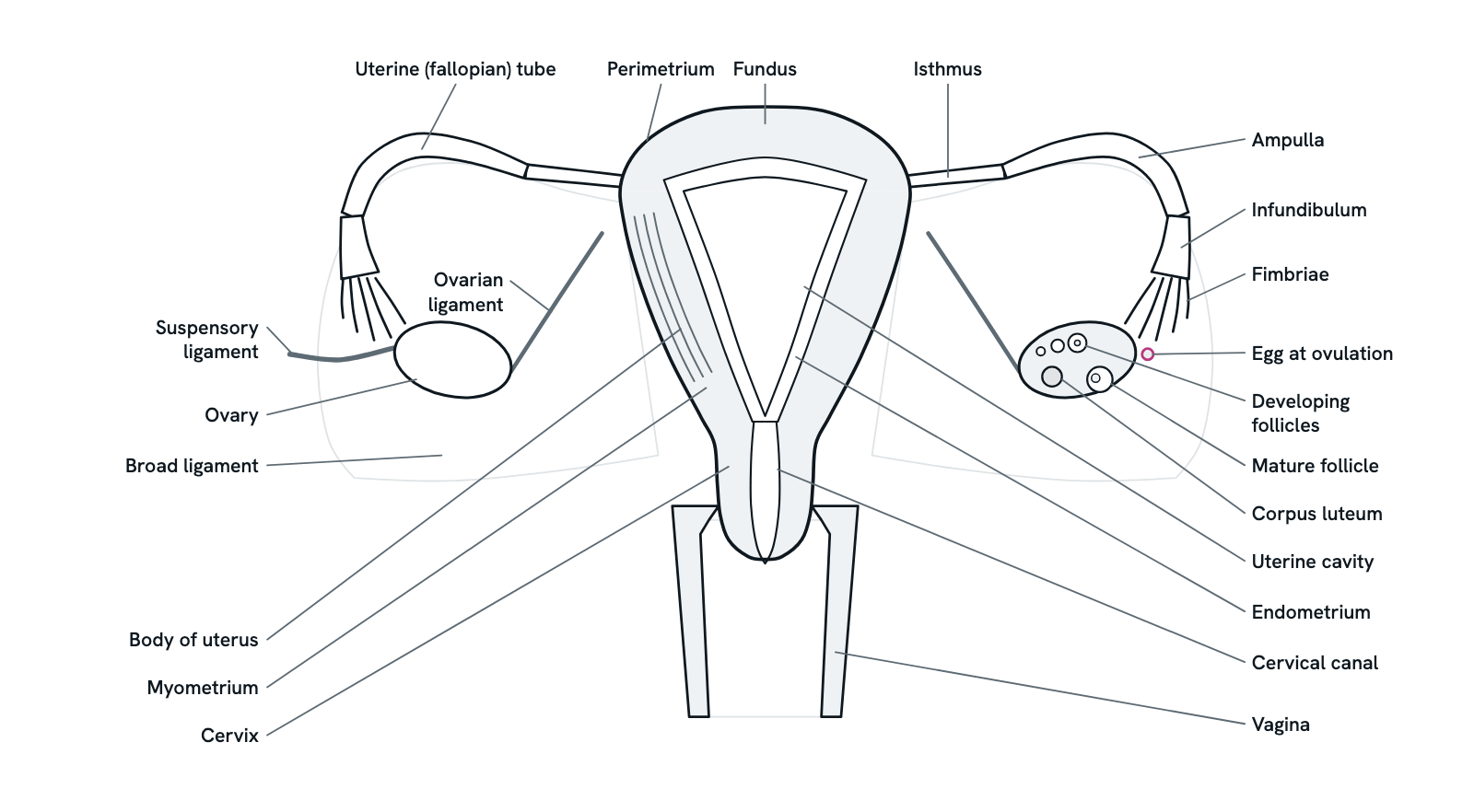
The internal organs of the female reproductive system seen from the front, as textbooks draw them: the uterus cut open to show its cavity and the layers of its wall, the two uterine (fallopian) tubes, the ovaries (one cut open to show its follicles) and the vagina below. Press any part to see what it is and what it does, or take the tour from the ovary to the vagina. Simplified, not to scale.
Also called Female reproductive organs, Uterus and ovaries or Female pelvic anatomy.
Press any part of the diagram, or pick one here, to see what it is and what it does.
Download the diagramChecked against its sources on 5 November 2026.
A tour
A tour of the female reproductive system
01The organs in order
The internal organs lie in the pelvis, seen here from the front. Two ovaries make eggs and hormones. Beside each, a uterine (fallopian) tube carries the egg towards the uterus, a hollow, pear-shaped muscular organ that lies behind the bladder and in front of the rectum. Its neck, the cervix, opens into the vagina, which leads to the outside. Follow an egg inwards: ovary, tube, uterus, cervix, vagina.
02The ovary: follicles, ovulation, corpus luteum
A female is born with all the primary oocytes she will have, each in a primordial follicle, arrested in prophase I of meiosis until puberty. Each cycle FSH drives a group of follicles to grow; one becomes dominant, and its granulosa cells make oestrogen. The LH surge ruptures it and the egg is released (ovulation). The remnant becomes the corpus luteum, which makes progesterone.
03The uterine tube and ectopic pregnancy
The fimbriae take up the egg, and cilia and muscle contractions carry it along the tube towards the uterus; fertilisation usually happens in the ampulla. From the uterus outwards, after a short part within the uterine wall, its parts are the isthmus, the ampulla and the infundibulum with its fimbriae. If the embryo implants outside the uterine cavity it is an ectopic pregnancy: about 1 to 2% of pregnancies in the US, 97% of them in the tube, most often in the ampulla (70%).
04The uterus and its three layers
From top to bottom the uterus has a fundus above the tubes, a body, a narrow isthmus and the cervix. Its wall has three layers: the perimetrium outside, the thick smooth-muscle myometrium, and the endometrium lining the cavity. A fertilised egg implants in the endometrium; the myometrium contracts in labour. In most women the uterus tilts forward over the bladder (anteverted) and its body bends forward on the cervix (anteflexed); in about 1 in 5 it tilts back (retroverted).
05The menstrual cycle in brief
Day 1 is the first day of bleeding, which lasts about 5 days. A cycle of 24 to 38 days is normal; 28 is typical. In the follicular phase, oestrogen from the growing follicle rebuilds the endometrium. Ovulation follows the LH surge around day 14, about 14 days before the next period. In the luteal phase, progesterone from the corpus luteum thickens the lining further. If no embryo implants, hormone levels fall and the lining is shed.
06The cervix
The cervix is the lower, narrow end of the uterus that connects it to the vagina; its canal runs from the internal os to the external os. In labour it thins and opens to 10 cm so the baby can pass. Long-lasting HPV infection causes almost all cervical cancers, so screening looks for HPV and for cell changes (a Pap or cytology test) before they become cancer.
07The vagina
The vagina is a fibromuscular tube about 10 cm long, lying between the bladder and the rectum, with the cervix projecting into its top. It carries menstrual flow out, receives the penis in intercourse and is the birth canal. Sperm deposited here swim up through the cervical canal into the uterus and the tubes, where they can live for 3 to 5 days; the egg lives only 12 to 24 hours.
08What holds the organs in place
The broad ligament, a double fold of peritoneum, hangs from the tubes like a sheet and holds the uterus, tubes and ovaries, with the uterine and ovarian vessels inside it. Each ovary is tied to the uterus by the ovarian ligament and to the pelvic side wall by the suspensory ligament, which carries the ovarian artery and vein. Here the suspensory ligament is drawn on the left only.
09The hormonal axis
The hypothalamus releases GnRH in pulses, which make the anterior pituitary secrete FSH and LH. FSH matures the follicles; LH triggers ovulation and keeps the corpus luteum making progesterone. Oestrogen from the follicle first holds LH down (negative feedback) and then, once high enough, drives the LH surge (positive feedback). If an embryo implants, its hCG keeps the corpus luteum going until the placenta takes over.
10Three common conditions
Endometriosis is endometrium-like tissue outside the uterine cavity, most often on the ovaries; it is oestrogen-dependent and causes chronic pelvic pain and reduced fertility. Fibroids are benign smooth-muscle tumours of the myometrium, the commonest gynaecological tumour, found in up to 70% of women by menopause. In polycystic ovary syndrome, high androgens and absent ovulation cause irregular or missed periods; it is the commonest cause of anovulatory infertility.
Parts
What each part of the female reproductive system does
- Ovary
- The female gonad: a paired, almond-sized organ beside the uterus. Its follicles hold the eggs and release one at ovulation; the follicles and the corpus luteum left after ovulation make the hormones oestrogen and progesterone.
- Developing follicles
- Each follicle is an egg (oocyte) wrapped in granulosa cells. A female is born with all her primary oocytes, arrested in prophase I of meiosis until puberty; from then on FSH drives a group of follicles to grow each cycle.
- Mature (Graafian) follicle
- The dominant follicle, with a fluid-filled antrum, bulging from the surface of the ovary just before ovulation. Its granulosa cells make most of the oestrogen of the follicular phase; the LH surge makes it rupture.
- Egg at ovulation
- The secondary oocyte released when the mature follicle ruptures, about 14 days before the next period. It lives about 12 to 24 hours unless fertilised, and is taken up by the fimbriae of the tube.
- Corpus luteum
- What is left of the follicle after ovulation. It secretes progesterone, which keeps the endometrium ready for implantation; without a pregnancy it degenerates after about 10 days into the corpus albicans, and the period follows.
- Uterine (fallopian) tube
- A paired tube from the ovary to the uterus, along which the egg is carried by cilia and muscle contractions and where fertilisation usually happens, most often in the ampulla. The tube on the right of the picture is labelled part by part.
- Isthmus (of the tube)
- The narrow medial part of the tube, next to the uterus; beyond it a short uterine part crosses the wall of the uterus to open into the cavity. About 12% of ectopic pregnancies implant here.
- Ampulla
- The long, wider middle part of the tube and the usual site of fertilisation. About 70% of ectopic pregnancies implant here, more than anywhere else.
- Infundibulum
- The funnel-shaped lateral end of the tube, open to the pelvic cavity near the ovary and fringed by the fimbriae.
- Fimbriae
- Finger-like fringes of the infundibulum that lie over the ovary and take up the egg released at ovulation, carrying it into the tube. About 11% of ectopic pregnancies implant on them.
- Ovarian ligament
- A fibrous cord from the medial pole of the ovary to the side of the uterus, just below where the tube enters. Also called the utero-ovarian ligament.
- Suspensory ligament of the ovary
- A fold of peritoneum from the lateral pole of the ovary to the pelvic side wall, carrying the ovarian artery and vein. Also called the infundibulopelvic ligament; drawn on the left only.
- Broad ligament
- A double fold of peritoneum draped over the tubes, from the sides of the uterus to the pelvic side walls. Its parts are the mesometrium, the mesosalpinx (round the tube) and the mesovarium (to the ovary); the uterine and ovarian vessels run in it.
- Fundus
- The broad, rounded top of the uterus, above the level at which the two tubes enter.
- Body of uterus
- The main part of the uterus, below the tubes, narrowing to the isthmus above the cervix. Its cavity, an inverted triangle in this section, is where the embryo implants and grows. The uterus is about 8 cm long, 5 cm wide and 4 cm thick.
- Perimetrium
- The outer coat of the uterus, a thin serous layer continuous with the peritoneum of the pelvis.
- Myometrium
- The thick middle layer of smooth muscle. Its contractions push the baby out in childbirth; fibroids (leiomyomas) are benign tumours that grow in it.
- Endometrium
- The mucous lining of the cavity: a thin basal layer and a thicker functional layer that thickens under oestrogen and progesterone each cycle, receives the embryo if one implants, and is shed at menstruation if not.
- Cervix
- The lower, narrow end of the uterus, projecting into the top of the vagina. In labour it thins and opens (dilates) to 10 cm. Cervical screening looks for HPV and for cell changes before they become cancer.
- Cervical canal
- The passage through the cervix, from the internal os at the uterine cavity to the external os in the vagina. Sperm pass up through it and menstrual blood comes down it.
- Vagina
- A fibromuscular tube about 10 cm long from the cervix to the outside, between the bladder in front and the rectum behind. It carries menstrual flow, receives the penis in intercourse and is the birth canal.
Dictionary
The terms to know
Practise it
The quizzes and cards that fit
- Human Anatomy quizzes
- Shoulder and Rotator Cuff15 questions
- Knee Joint15 questions
- Arteries and Veins15 questions
- Physiology quizzes
- Exercise Physiology15 questions
- Puberty15 questions
- Blood Clotting15 questions
- Medicine quizzes
- Labor and Delivery15 questions
- IV Fluids15 questions
- Sepsis15 questions
- Human Anatomy flashcards
The subject's terms as a ready-made deck, free to flip through.
Next to it
More diagrams
Sources
Where the facts come from
- StatPearls: Physiology, Uterus (NCBI Bookshelf)
- StatPearls: Anatomy, Abdomen and Pelvis: Uterus (NCBI Bookshelf)
- StatPearls: Anatomy, Abdomen and Pelvis, Ovary (NCBI Bookshelf)
- StatPearls: Anatomy, Abdomen and Pelvis: Fallopian Tube (NCBI Bookshelf)
- StatPearls: Anatomy, Abdomen and Pelvis: Broad Ligaments (NCBI Bookshelf)
- StatPearls: Physiology, Ovulation (NCBI Bookshelf)
- StatPearls: Physiology, Menstrual Cycle (NCBI Bookshelf)
- StatPearls: Ectopic Pregnancy (NCBI Bookshelf)
- StatPearls: Endometriosis (NCBI Bookshelf)
- StatPearls: Fertility Sparing Management in Uterine Fibroids (NCBI Bookshelf)
- Bouyer and others, Sites of ectopic pregnancy: a 10 year population-based study of 1800 cases, Human Reproduction 2002
- Office on Women's Health: Your menstrual cycle
- NICHD: About Menstruation
- NICHD: About Endometriosis
- NICHD: About Uterine Fibroids
- NICHD: About Polycystic Ovary Syndrome
- NICHD: What are the stages of labor?
- NCI SEER Training Modules: Female Reproductive System
- National Cancer Institute: Cervical Cancer
- CDC: Screening for Cervical Cancer
Drawn and written by biofideX from these sources.