Male Reproductive System Diagram
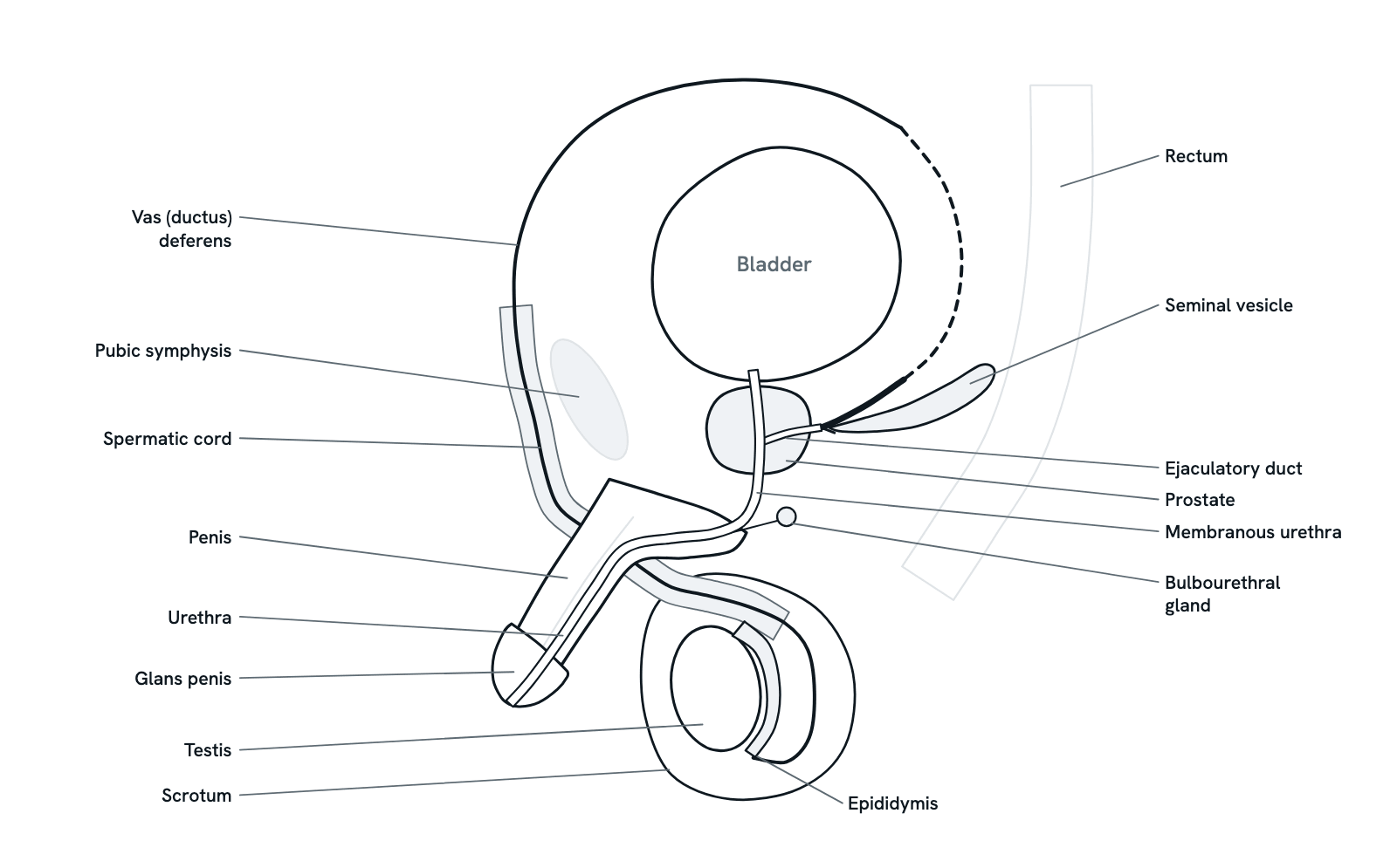
The male reproductive organs from the side: testis, epididymis, vas deferens, the glands that make semen, the prostate, urethra and penis.
A section through the male pelvis seen from the side, facing left, as textbooks draw it: the bladder with the prostate below it, the seminal vesicle behind, the vas deferens looping from the testis over the bladder to the ejaculatory duct, and the urethra through the prostate and along the penis. Press any part to see what it is and what it does, or take the tour along the path of a sperm. Simplified, not to scale.
Also called Male reproductive organs or Male pelvic anatomy.
Press any part of the diagram, or pick one here, to see what it is and what it does.
Download the diagramChecked against its sources on 5 November 2026.
A tour
A tour of the male reproductive system
01The organs in order
Seen from the side, the testis hangs in the scrotum below the pelvis with the epididymis along its back. The vas deferens climbs from there into the pelvis, loops over the bladder and comes down behind it, where the seminal vesicle joins it. The prostate sits under the bladder round the urethra, which then runs along the penis. The rectum lies behind all of these.
02The path of a sperm: SEVEN UP
Sperm are made in the seminiferous tubules of the testis, mature and wait in the epididymis, and at ejaculation travel up the vas deferens, through the ejaculatory duct (where the seminal vesicle's fluid joins, and which runs through the prostate) and out along the urethra and penis. The mnemonic SEVEN UP keeps the order: Seminiferous tubules, Epididymis, Vas deferens, Ejaculatory duct, Nothing, Urethra, Penis.
03The testis and the scrotum
Inside each testis, coiled seminiferous tubules make sperm from puberty onwards, a process of about 74 days: spermatogonia multiply, their daughter cells halve their chromosomes by meiosis and are shaped into motile sperm, nursed by Sertoli cells in the tubule wall. Between the tubules, Leydig cells make testosterone. The scrotum holds the testes about 3 degrees C below body temperature, which viable sperm need.
04The epididymis: maturing and storing sperm
Sperm leave the testis immature and unable to fertilise an egg. They pass into the head of the epididymis, a single tube coiled so tightly that it would stretch about 6 metres, and mature as they move down its body; mature sperm are stored in its tail, where the vas deferens begins.
05The vas deferens and vasectomy
At ejaculation the muscular wall of the vas deferens drives sperm up through the spermatic cord and the inguinal canal, over the bladder and down behind it to the ampulla. A vasectomy, the only permanent male sterilisation, cuts and ties the vas on each side; it succeeds in about 99.7% of cases, with complications in 1 to 2%.
06The glands that make semen
Semen is sperm plus the secretions of three glands. The seminal vesicles add about 60 to 70% of the volume: a thick fluid with fructose to feed the sperm, prostaglandins and proteins that clot the semen briefly. The prostate adds most of the rest, a thin, milky, alkaline fluid that protects sperm from the acid of the vagina. The bulbourethral glands add a little alkaline mucus. One ejaculate is 1.5 to 6 mL with about 50 to 150 million sperm per mL.
07The prostate, the urethra and BPH
The urethra carries urine and semen through the prostate, the pelvic floor and the penis. With age the prostate's transition zone, which surrounds the urethra, enlarges (benign prostatic hyperplasia): it squeezes the urethra, the bladder has to work harder, and in time emptying may be incomplete. At autopsy it is found in 50 to 60% of men in their sixties and 80 to 90% over 70. PSA, a protein made by prostate cells, rises in cancer but also in BPH and prostatitis.
08Erection and ejaculation
Erection follows parasympathetic signals that relax the smooth muscle of the penile arteries and erectile tissue, so the corpora cavernosa and corpus spongiosum fill with blood. Ejaculation has two phases: emission, in which sympathetic nerves contract the vas deferens, seminal vesicles and prostate and close the bladder neck so semen collects in the urethra, then expulsion by rhythmic contractions of the pelvic floor. Hence: parasympathetic to point, sympathetic to shoot.
09The hormonal axis
The hypothalamus releases GnRH, which makes the anterior pituitary secrete LH and FSH. LH acts on the Leydig cells to make testosterone, which drives spermatogenesis and the male secondary sexual characteristics; FSH acts on the Sertoli cells to support spermatogenesis. Testosterone feeds back to hold LH down, and the Sertoli cells' inhibin B does the same for FSH.
Parts
What each part of the male reproductive system does
- Testis
- The male gonad, an oval gland about 3 to 5 cm long and 2 to 3 cm wide, held in the scrotum by the spermatic cord. Its seminiferous tubules make sperm and its Leydig cells make testosterone; the left usually hangs a little lower than the right.
- Epididymis
- A tightly coiled tube, about 6 metres long if straightened, lying along the back of the testis as a head, body and tail. Sperm leaving the testis cannot yet fertilise an egg; they mature as they pass along it and are stored in its tail.
- Scrotum
- The sac of skin and muscle below the penis that holds the testes, one in each compartment. It keeps them about 3 degrees C cooler than the body, which sperm production needs.
- Vas (ductus) deferens
- A muscular tube that carries sperm from the tail of the epididymis up through the inguinal canal in the spermatic cord, over the bladder and down behind it, where its widened end, the ampulla, meets the duct of the seminal vesicle. A vasectomy cuts and ties it.
- Spermatic cord
- The bundle that suspends the testis: the vas deferens, the testicular artery, veins and nerves and the cremaster muscle, wrapped in fascia, running from the scrotum through the inguinal canal into the abdomen.
- Seminal vesicle
- A paired coiled gland, 3 to 5 cm long, behind the base of the bladder and above the prostate. Its thick fluid, rich in fructose and prostaglandins, makes about 60 to 70% of semen; its duct joins the vas deferens to form the ejaculatory duct.
- Ejaculatory duct
- The short duct formed where the ampulla of the vas deferens meets the duct of the seminal vesicle. It passes through the prostate and opens into the prostatic urethra at the verumontanum.
- Prostate
- A firm, walnut-sized gland just below the bladder, encircling the urethra where it leaves the bladder. Its thin, milky, alkaline fluid protects sperm from the acid of the vagina and helps them move; it makes up most of the rest of semen after the seminal vesicles.
- Bulbourethral (Cowper's) gland
- A paired pea-sized gland behind the membranous urethra, near the base of the penis. Before ejaculation it secretes an alkaline mucus into the start of the spongy urethra, which neutralises any acid and lubricates the tip of the penis.
- Urethra
- The final path for both urine and semen. It runs from the bladder neck through the prostate (prostatic part), through the pelvic floor (membranous part) and along the penis (spongy part) to the opening at the tip of the glans.
- Penis
- Three columns of erectile tissue: two corpora cavernosa on top and the corpus spongiosum below, which carries the urethra and expands at the end into the glans. It has a root, fixed to the pubic arch, a body and the glans.
- Glans penis
- The expanded end of the corpus spongiosum at the tip of the penis. The urethra opens at its tip (the meatus); a loose fold of skin, the prepuce or foreskin, covers it.
- Bladder
- Stores the urine that comes down the ureters from the kidneys, for orientation here: the prostate sits below its neck, and the seminal vesicles lie behind its base. Urine and semen share the urethra below it.
- Pubic symphysis
- The slightly movable joint at the front of the pelvis where the two pubic bones meet, with a pad of fibrocartilage between them. The bladder sits just behind it. Drawn faintly for orientation.
- Rectum
- The last part of the large bowel, behind the bladder, the seminal vesicles and the prostate, from which a layer of fascia separates it. Drawn faintly for orientation.
Dictionary
The terms to know
Practise it
The quizzes and cards that fit
- Human Anatomy quizzes
- Shoulder and Rotator Cuff15 questions
- Knee Joint15 questions
- Arteries and Veins15 questions
- Physiology quizzes
- Exercise Physiology15 questions
- Puberty15 questions
- Blood Clotting15 questions
- Medicine quizzes
- Labor and Delivery15 questions
- IV Fluids15 questions
- Sepsis15 questions
- Human Anatomy flashcards
The subject's terms as a ready-made deck, free to flip through.
Next to it
More diagrams
Sources
Where the facts come from
- StatPearls: Anatomy, Abdomen and Pelvis: Testes (NCBI Bookshelf)
- StatPearls: Anatomy, Abdomen and Pelvis, Scrotum (NCBI Bookshelf)
- StatPearls: Anatomy, Abdomen and Pelvis, Male Genitourinary Tract (NCBI Bookshelf)
- StatPearls: Anatomy, Abdomen and Pelvis, Seminal Vesicle (NCBI Bookshelf)
- StatPearls: Anatomy, Abdomen and Pelvis, Prostate (NCBI Bookshelf)
- StatPearls: Anatomy, Abdomen and Pelvis, Penis (NCBI Bookshelf)
- StatPearls: Anatomy, Abdomen and Pelvis: Bladder (NCBI Bookshelf)
- StatPearls: Anatomy, Abdomen and Pelvis: Inguinal Region (Inguinal Canal) (NCBI Bookshelf)
- StatPearls: Histology, Spermatogenesis (NCBI Bookshelf)
- StatPearls: Histology, Sertoli Cell (NCBI Bookshelf)
- StatPearls: Histology, Leydig Cells (NCBI Bookshelf)
- StatPearls: Physiology, Testosterone (NCBI Bookshelf)
- StatPearls: Physiology, Luteinizing Hormone (NCBI Bookshelf)
- StatPearls: Vasectomy (NCBI Bookshelf)
- StatPearls: Benign Prostatic Hyperplasia (NCBI Bookshelf)
- NCI SEER Training Modules: Male Reproductive System (testes, duct system, accessory glands, penis, sexual response and hormone control)
- NCI SEER Training Modules: Articulations (the symphysis pubis)
- NIDDK: Enlarged Prostate (Benign Prostatic Hyperplasia)
- National Cancer Institute: Prostate-Specific Antigen (PSA) Test
- StatPearls: Physiology, Erection (NCBI Bookshelf)
- StatPearls: Premature Ejaculation (NCBI Bookshelf; the phases of normal ejaculation)
- Giuliano, Neurophysiology of erection and ejaculation, Journal of Sexual Medicine 2011
Drawn and written by biofideX from these sources.