Meiosis Diagram
The stages of meiosis I and II: how one diploid cell becomes four haploid, genetically different gametes, through crossing over and two divisions.
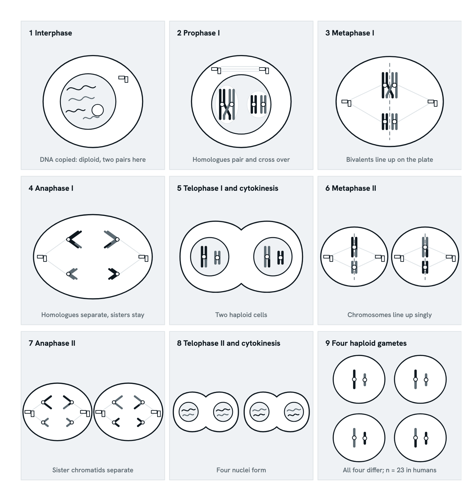
Meiosis makes eggs and sperm. One round of DNA replication is followed by two divisions, so one diploid cell gives four haploid cells, each with one chromosome of every pair: 23 in humans instead of 46. The cell here has two pairs of homologous chromosomes, one long and one short; of each pair, the chromosome from one parent is in ink and the other in grey, so you can follow each one.
Also called Stages of meiosis, Meiosis I and II or Meiosis 1 and 2.
Step 1 of 10
Meiosis at a glance
Meiosis makes gametes. After a single round of DNA replication the cell divides twice, so one diploid cell becomes four haploid cells, each with one chromosome of every pair: 23 in humans instead of 46. On the way, crossing over and the random sorting of the pairs make every gamete genetically different. The cell here has two pairs of homologous chromosomes, one long and one short.
Use ← and → to move between steps.
Step 1 of 10: Meiosis at a glance
Download the diagramChecked against its sources on 5 November 2026.
Step by step
The steps of the Meiosis
01Meiosis at a glance
Meiosis makes gametes. After a single round of DNA replication the cell divides twice, so one diploid cell becomes four haploid cells, each with one chromosome of every pair: 23 in humans instead of 46. On the way, crossing over and the random sorting of the pairs make every gamete genetically different. The cell here has two pairs of homologous chromosomes, one long and one short.
02Interphase: the DNA is copied
The cell is diploid: it has two of every chromosome, one from each parent, drawn here in ink and in grey. In S phase every chromosome is copied, so each enters meiosis as two identical sister chromatids held together by cohesin. This is the only replication: both divisions share out these copies, and it is that which halves the number.
03Prophase I: pairing and crossing over
The longest stage, with sub-stages of its own (leptotene, zygotene, pachytene, diplotene and diakinesis). Each chromosome pairs with its homologue along its whole length, held by a protein scaffold, the synaptonemal complex: the pair is a bivalent, or tetrad of four chromatids. In pachytene two non-sister chromatids break and rejoin, swapping segments; from diplotene this crossing over shows as a chiasma. Almost every pair gets at least one, usually only a few.
04Metaphase I: bivalents on the plate
Bivalents, not single chromosomes, line up on the metaphase plate, each held together by its chiasmata. The two sister kinetochores of one homologue attach to fibres from the same pole, and the other homologue faces the opposite pole. Which way round each pair lies is random and independent of the other pairs: Mendel's independent assortment, the second source of new combinations.
05Anaphase I: the homologues separate
Cohesin along the chromosome arms is cut, the chiasmata come apart and the homologues are pulled to opposite poles. The sister chromatids of each stay joined, because the cohesin at their centromeres is protected by the protein shugoshin. This is the reductional division: each pole now has one chromosome of every pair, so the number is halved, and here the long chromosome in ink travels with the short one in grey.
06Telophase I and cytokinesis: two haploid cells
The cell divides into two haploid cells. Each has one homologue of every pair, still as two chromatids, and the two cells differ: on the left the long chromosome came from one parent and the short one from the other. There is no S phase before the second division; without that omission the chromosome number could not be halved.
07Metaphase II: like a mitosis
Meiosis II runs like a mitosis in each of the two cells. The chromosomes line up singly on a new metaphase plate, and now the two sister kinetochores of each attach to opposite poles, exactly as they do in mitosis.
08Anaphase II and telophase II: the sisters separate
The cohesin that was left at the centromeres is cut, and the sister chromatids separate and move to opposite poles: the equational division, which leaves the number of chromosomes as it was. A nuclear envelope forms round each of the four sets, and cytokinesis divides each cell in two.
09Four different gametes
Four haploid gametes, with 23 chromosomes each in humans. No two are the same: crossing over has made recombinant chromosomes, part ink and part grey, and independent assortment has dealt the homologues of the two pairs in different combinations. In women only one of the four becomes the egg; the others are polar bodies. At fertilisation an egg's 23 chromosomes join a sperm's 23, and the new cell has 46.
10Compared with mitosis, and when it goes wrong
Mitosis gives two identical diploid cells, meiosis four different haploid ones. Three things set meiosis I apart: homologues pair and cross over, sister kinetochores attach to the same pole, and cohesin is removed in two steps, arms first and centromeres later. When chromosomes fail to separate (nondisjunction) a gamete gets the wrong number: about 20% of human eggs do, far more than sperm, and an extra chromosome 21, usually from the egg, causes Down syndrome.
Parts
What each part of the meiosis does
- Interphase
- Before meiosis the diploid cell copies its DNA in S phase, so every chromosome enters meiosis as two identical sister chromatids. There is no further replication between the two divisions.
- Prophase I
- The longest stage. Homologous chromosomes pair along their length as bivalents, and non-sister chromatids exchange segments by crossing over, seen as chiasmata.
- Metaphase I
- The bivalents line up on the metaphase plate, each homologue facing one pole. Which way round a pair lies is random and independent of the other pairs.
- Anaphase I
- The homologues of each pair are pulled to opposite poles, while the sister chromatids stay together: the reductional division, which halves the chromosome number.
- Telophase I and cytokinesis
- The cell divides into two haploid cells. Each has one chromosome of every pair, still as two chromatids, and goes straight on to meiosis II without copying its DNA.
- Metaphase II
- In each cell the chromosomes line up singly on a new metaphase plate, with the sister kinetochores now facing opposite poles, as in mitosis.
- Anaphase II
- The cohesin still holding the centromeres is cut and the sister chromatids separate to opposite poles: the equational division, which leaves the chromosome number unchanged.
- Telophase II and cytokinesis
- A nuclear envelope forms round each of the four sets of chromosomes, and each of the two cells divides in two.
- Haploid gametes
- The four cells that result, each with one chromosome of every pair, 23 in humans, and each with a different combination. In men all four become sperm; in women only one becomes the egg and the others are polar bodies. An egg and a sperm fuse at fertilisation to restore the diploid 46.
- Homologous chromosomes
- The two chromosomes of a pair, one from each parent, carrying the same genes in the same order, though not always the same alleles. One is drawn in ink and the other in grey.
- Sister chromatids
- The two identical copies of one chromosome made in S phase, joined by cohesin. They stay together through meiosis I and separate only in anaphase II.
- Chiasma (crossing over)
- The cross-shaped point where two non-sister chromatids of a pair have broken and rejoined, swapping segments. Chiasmata also hold the homologues together until anaphase I.
- Bivalent (tetrad)
- A pair of homologous chromosomes paired in prophase I, four chromatids in all, held first by the synaptonemal complex and then by its chiasmata. Almost every one has at least one crossover.
- Spindle fibres
- Microtubules from the two poles that attach to the kinetochores and pull chromosomes apart: whole homologues in meiosis I, sister chromatids in meiosis II.
- Centrosome
- The organiser of the spindle's microtubules at each pole. Oocytes are an exception: their spindle assembles round the chromosomes without centrosomes.
- Metaphase plate
- The plane midway between the poles where the bivalents line up in metaphase I and the single chromosomes in metaphase II. It is a position, not a structure.
- Nuclear envelope
- The double membrane round the nucleus. It breaks up before each division, so the spindle can reach the chromosomes, and re-forms round each new set.
- Centromere
- The constricted region where the sister chromatids are held together and where the kinetochores form. In meiosis I both sister kinetochores attach to the same pole.
- Nucleolus
- The part of the nucleus that makes ribosomes. It fades as the chromosomes condense in prophase I.
- The cell
- The diploid cell dividing here, then the two haploid cells of meiosis II. The nine panels are not to the same scale.
Dictionary
The terms to know
Practise it
The quizzes and cards that fit
- Genetics quizzes
- Incomplete Dominance and Codominance15 questions
- Pedigree Analysis15 questions
- Blood Types15 questions
- Cell Biology quizzes
- Characteristics of Life15 questions
- Lab Safety15 questions
- Cell Theory15 questions
- Genetics flashcards
The subject's terms as a ready-made deck, free to flip through.
- Dihybrid Cross Calculator
The Punnett square for any cross of two genes, with the gametes, the genotype and phenotype ratios, gene interactions and a chi-square test of your counts.
Next to it
More diagrams
Sources
Where the facts come from
- Ohkura, Meiosis: an overview of key differences from mitosis, Cold Spring Harbor Perspectives in Biology 2015 (PMC)
- Jones, Kleckner and Zickler, Meiosis through three centuries, Chromosoma 2024 (PMC)
- NHGRI Talking Glossary: Meiosis
- NHGRI Talking Glossary: Crossing Over
- Nagaoka, Hassold and Hunt, Human aneuploidy: mechanisms and new insights into an age-old problem, Nature Reviews Genetics 2012 (PMC)
- MedlinePlus Genetics: Down syndrome
Drawn and written by biofideX from these sources.