Kidney Diagram
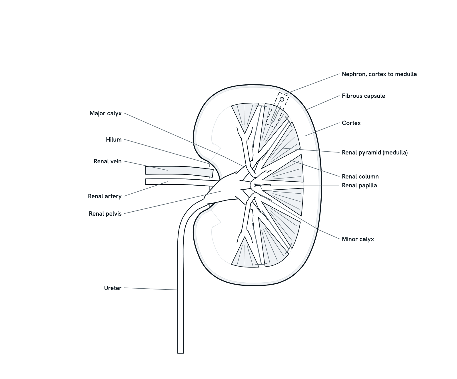
The kidney cut lengthways: capsule, cortex and medulla, the pyramids and their papillae, the calyces, pelvis and ureter, and the vessels at the hilum.
A kidney in coronal section, the hilum facing the midline. Blood arrives by the renal artery, is filtered in about a million nephrons across the cortex and medulla, and the urine drains from the tips of the pyramids through the calyces and renal pelvis into the ureter. Press any part for what it is and what it does, or take the tour. Simplified, not to scale.
Also called Kidney anatomy, Renal anatomy or Kidney cross section.
Press any part of the diagram, or pick one here, to see what it is and what it does.
Download the diagramChecked against its sources on 5 November 2026.
A tour
A tour of the kidney
01The kidneys at a glance
The two kidneys are bean-shaped organs about the size of a fist, each roughly 12 cm long, 6 cm wide and 3 cm thick. They lie against the back wall of the abdomen behind the peritoneum, so they are retroperitoneal, between the twelfth thoracic and third lumbar vertebrae and protected by the lower ribs. The right kidney sits a little lower than the left because the liver pushes it down. This is a kidney cut in the coronal plane, its hilum facing the midline.
02Capsule, hilum and sinus
A tough fibrous capsule closely wraps each kidney and supports the soft tissue inside; outside it lie a thick layer of fat and the renal fascia, which hold the kidney in place. On the medial side is an indentation, the hilum, where the renal artery enters and the renal vein and ureter leave. The hilum opens into a cavity inside the kidney, the renal sinus, which holds the renal pelvis, the calyces and the first branches of the vessels.
03Cortex and medulla
Under the capsule lies the cortex, the outer reddish layer, and inside it the darker medulla, made up of cone-shaped renal pyramids. Cortex also runs down between the pyramids as the renal columns. The cortex holds the glomeruli, where the blood is filtered, and the coiled tubules; the pyramids look striped because straight tubules and blood vessels run through them, and the medulla's main work is concentrating the urine.
04Pyramids and papillae
Each pyramid has its wide base against the cortex and its pointed tip, the renal papilla, aimed at the sinus. The collecting ducts open at the papilla, and the urine passes into the cup-shaped minor calyx that surrounds it. A minor calyx may be simple, draining one papilla, or compound, draining two or three; 70% of kidneys have 7 to 9 of them.
05The collecting system
Minor calyces converge into major calyces, usually an upper and a lower group, sometimes a third for the middle as drawn here; in some kidneys a minor calyx drains straight into the renal pelvis. The major calyces open into the funnel of the renal pelvis, which narrows at the hilum into the ureter. So urine made in the nephrons passes through the collecting ducts, the calyces and the pelvis, then down the ureter, a thin tube of muscle, to the bladder.
06Blood supply
The kidneys receive 20 to 25% of the heart's output. Usually a single renal artery enters at the hilum and divides in the sinus into five segmental end arteries. Interlobar arteries run out between the pyramids in the renal columns, arcuate arteries curve along the base of the medulla, and interlobular arteries radiate towards the capsule, giving off the afferent arterioles to the glomeruli. The veins follow the same route back to the renal vein, but connect between segments.
07A million nephrons
Each kidney holds about a million nephrons, the units that make urine: on average 900,000 to 1 million, though single kidneys range from about 200,000 to more than 2.5 million, and no new ones form after about 36 weeks of gestation. The glomeruli and coiled tubules sit in the cortex; the loops of Henle and collecting ducts run down through the pyramids, so one nephron spans cortex and medulla, as the dashed box shows.
08Filtering the blood
In each nephron the glomerulus lets water, wastes and other small molecules pass into the tubule while proteins and blood cells stay in the blood; the tubule then returns almost all the water, with the minerals and nutrients the body needs, and removes extra acid. Healthy kidneys filter about half a cup of blood every minute, around 150 quarts (140 litres) a day, of which only 1 to 2 quarts become urine.
09More than urine
The kidneys also balance water, salts and minerals such as sodium, potassium, calcium and phosphorus, remove acid made by the body's cells, and make hormones. Granular cells of the juxtaglomerular apparatus, beside each glomerulus, release renin, which helps regulate blood pressure. The kidneys make erythropoietin, the hormone that tells the bone marrow to make red blood cells, and they turn vitamin D into its active form, calcitriol, which the bones need.
10Stones and chronic kidney disease
Kidney stones form when certain minerals are high in the urine, most often as calcium oxalate. A small stone passes with little or no pain; a larger one may get stuck in the ureter. About 11% of men and 6% of women in the US have had one. Chronic kidney disease, damage that stops the kidneys filtering as they should, is tracked by the glomerular filtration rate, estimated from blood creatinine: a GFR below 60 may mean kidney disease, 15 or less is kidney failure.
Parts
What each part of the kidney does
- Fibrous capsule
- A tough, fibrous coat of connective tissue that closely envelops the kidney and supports the soft tissue inside. Outside it lies a thick layer of fat, and the renal fascia holds the kidney in place.
- Renal cortex
- The outer, reddish layer next to the capsule. It holds the glomeruli, where the blood is filtered, with their afferent and efferent arterioles, and the coiled tubules of the nephrons wrapped in peritubular capillaries.
- Renal medulla: the pyramids
- The inner, darker region, made of cone-shaped renal pyramids with their bases against the cortex. They look striped because straight tubules and blood vessels run through them; the loops of Henle and collecting ducts pass down here, and the medulla's main job is concentrating the urine.
- Renal column
- A portion of cortex that extends down between two neighbouring pyramids. The interlobar arteries and veins run through the columns on their way to and from the cortex.
- Renal papilla
- The pointed tip of a pyramid, directed towards the centre of the kidney. The collecting ducts open here, and the urine passes from them into the minor calyx that surrounds the papilla.
- Minor calyx
- A cup that surrounds the papilla of a pyramid and collects the urine from it. A minor calyx may be simple, draining one papilla, or compound, draining two or three; 70% of kidneys have 7 to 9 minor calyces.
- Major calyx
- Several minor calyces converge to form a major calyx. Most kidneys have two main calyceal groups, upper and lower, and some a third for the middle of the kidney; here a middle group drains the two lateral pyramids.
- Renal pelvis
- The funnel-shaped cavity that collects urine from the major calyces and is continuous with the ureter. Its place varies: it may lie inside the renal sinus, outside the kidney beyond the hilum, or partly in each.
- Ureter
- The thin muscular tube that carries urine from the renal pelvis down to the bladder, one from each kidney. A kidney stone too large to pass can get stuck in it.
- Hilum
- The indentation on the medial side of the kidney, where the renal artery enters and the renal vein and ureter leave. It leads into a cavity inside the kidney, the renal sinus, which holds the pelvis, the calyces and the first branches of the vessels.
- Renal artery
- Usually one to each kidney; together the kidneys get 20 to 25% of cardiac output. In the sinus it divides into five segmental end arteries; interlobar arteries run between the pyramids, arcuate arteries curve along the base of the medulla, and interlobular arteries reach the cortex and give off afferent arterioles.
- Renal vein
- Carries the filtered blood out of the kidney at the hilum. The veins follow the same pattern as the arteries, but unlike the segmental arteries, veins from different segments connect with one another.
- Nephron
- The kidney's filtering unit, about a million to a kidney. Its glomerulus filters the blood in the cortex and its tubule returns what the body needs; its loop of Henle, and the collecting duct it drains into, run down into the medulla. See the nephron diagram for how it works.
Dictionary
The terms to know
Practise it
The quizzes and cards that fit
- Human Anatomy quizzes
- Shoulder and Rotator Cuff15 questions
- Knee Joint15 questions
- Arteries and Veins15 questions
- Physiology quizzes
- Exercise Physiology15 questions
- Puberty15 questions
- Blood Clotting15 questions
- Medicine quizzes
- Labor and Delivery15 questions
- IV Fluids15 questions
- Sepsis15 questions
- Human Anatomy flashcards
The subject's terms as a ready-made deck, free to flip through.
- Normal Lab Values
Typical adult ranges for blood, urine and spinal fluid tests, in conventional and SI units, with a source for every value.
Next to it
More diagrams
Sources
Where the facts come from
- StatPearls: Anatomy, Abdomen and Pelvis: Kidneys (NCBI Bookshelf)
- NIDDK: Your Kidneys and How They Work
- NCI SEER Training: Kidneys
- Russell and others, Renal Lymphatics: Anatomy, Physiology, and Clinical Implications, Frontiers in Physiology, 2019 (PubMed Central)
- Dumas and others, Phenotypic diversity and metabolic specialization of renal endothelial cells, Nature Reviews Nephrology, 2021 (PubMed Central)
- Morais and others, Kidney collecting system anatomy applied to endourology, International Brazilian Journal of Urology, 2024 (PubMed Central)
- Bertram and others, Human nephron number: implications for health and disease, Pediatric Nephrology, 2011 (PubMed)
- Peti-Peterdi and Harris, Macula Densa Sensing and Signaling Mechanisms of Renin Release, Journal of the American Society of Nephrology, 2010 (PubMed Central)
- NIDDK: Anemia in Chronic Kidney Disease
- NIDDK: Mineral and Bone Disorder in Chronic Kidney Disease
- NIDDK: Chronic Kidney Disease Tests and Diagnosis
- NIDDK: Kidney Stones, Definition and Facts
Drawn and written by biofideX from these sources.