Cell Membrane Diagram
The cell membrane in cross-section: the phospholipid bilayer, its proteins, cholesterol and sugar coat, and what each part does.
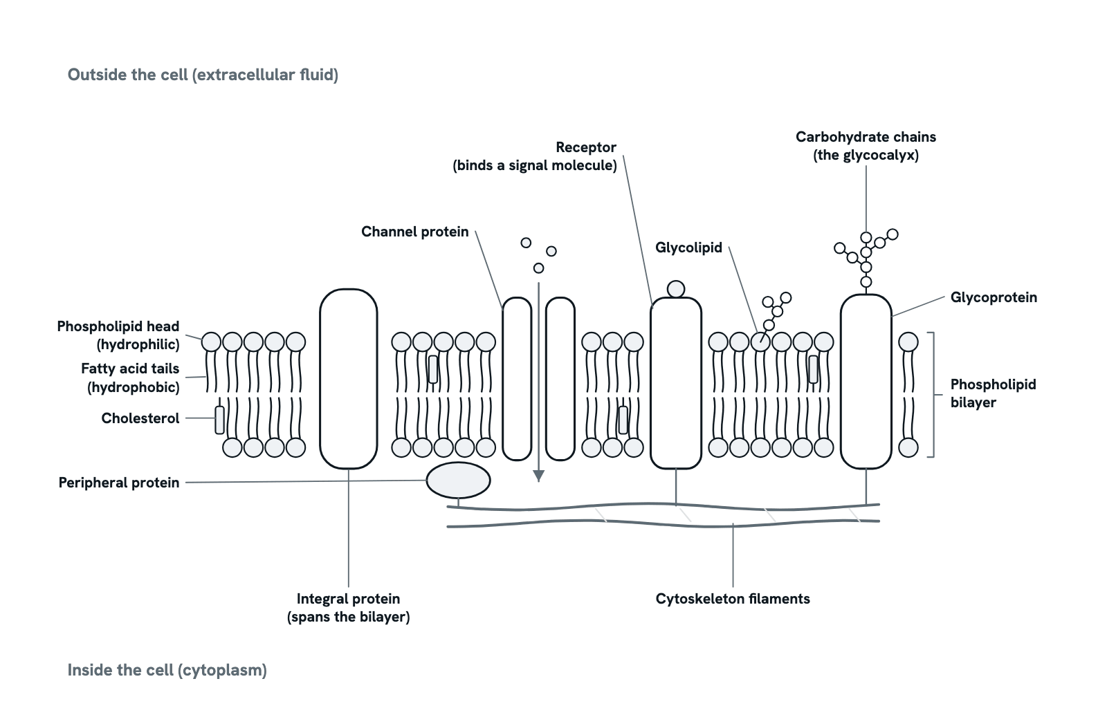
A cross-section of the plasma membrane as the fluid mosaic model describes it, the outside of the cell at the top. Two layers of phospholipids make a sheet a few nanometres thick; proteins span it or rest on it, cholesterol stiffens it, and sugar chains coat its outer face. Press any part to see what it is and what it does, or take the tour in order. Not to scale.
Also called Plasma membrane, Fluid mosaic model or Phospholipid bilayer.
Press any part of the diagram, or pick one here, to see what it is and what it does.
Download the diagramChecked against its sources on 5 November 2026.
A tour
A tour of the cell membrane
01The cell membrane at a glance
Every cell is wrapped in a membrane only a few nanometres thick: a double layer of phospholipids with proteins set in it and sugar chains on its outer face. It keeps the inside of the cell apart from the fluid outside, lets through what the cell needs and passes signals in. Singer and Nicolson named this arrangement the fluid mosaic model in 1972.
02The bilayer, and why it forms
A phospholipid is amphipathic: its phosphate head mixes with water, its two fatty acid tails do not. In water such lipids arrange themselves, without any help, into a sheet two molecules thick, heads outwards to the water on both faces and tails inwards, away from it. The hydrophobic core is about 3 nm thick, and the two leaflets differ in which lipids they hold.
03What crosses on its own
The oily core lets small uncharged molecules slip through by simple diffusion, water among them, and lipid-soluble molecules such as the steroid hormones, which reach receptors inside the cell. Ions and large polar molecules such as sugars, amino acids and the hormone insulin cannot cross a bare bilayer. So the membrane is selectively permeable, and its proteins decide what else gets in or out.
04Channels and carriers: facilitated diffusion
A channel protein is a water-filled pore through the membrane. Ions pass down their electrochemical gradient at no cost in energy, and the pore's size and charge pick which ion. Channels open and shut: to a ligand, to a change in voltage or to stretch. A carrier protein binds its passenger, a sugar for instance, changes shape and lets it go on the other side, down its gradient and more slowly than a channel. Both are passive transport.
05Pumps: active transport
Moving a solute against its gradient takes energy: from ATP, from light, or from the downhill flow of another ion through the same carrier. The sodium-potassium pump, an integral protein like this one, splits ATP to push three sodium ions out of the cell and bring two potassium ions in. In the brain's grey matter these pumps use about three quarters of the energy. The sodium gradient they build then drives glucose and amino acids into cells by symport.
06Cholesterol and fluidity
Lipids and proteins drift sideways within their leaflet, which is what makes the membrane fluid; flipping from one leaflet to the other is rare, so the two faces stay different. Cholesterol, a stiff four-ring sterol, sits among the tails of animal cell membranes, and the plasma membrane holds more of it than any other membrane in the cell. It makes the membrane thicker and more rigid, and gathers with sphingolipids into small rafts, 10 to 200 nm across.
07The glycocalyx: sugars on the outside
Sugar chains are attached to many membrane proteins and some lipids, always on the outer face, and with the longer chains of proteoglycans they form the glycocalyx, a gel-like coat that every cell in the body carries. The chains are built in the endoplasmic reticulum and the Golgi. The coat protects the cell and is a barrier to pathogens, keeps white cells from sticking where they should not, and gives each cell a surface that other cells and the immune system recognise.
08Receptors and signalling
Water-soluble signals such as insulin and adrenaline cannot enter the cell, so they bind receptors in the membrane instead. A receptor spanning the bilayer binds its signal on the outside and changes on the inside: the adrenaline receptor passes the message to a G protein, which switches on an enzyme, and the nicotinic acetylcholine receptor is itself a channel that opens to let sodium in. About a quarter of all drugs act on G-protein-coupled receptors.
09The fluid mosaic model
In 1972 Singer and Nicolson described the membrane as a two-dimensional solution: globular integral proteins, amphipathic like the lipids, afloat in a fluid phospholipid bilayer, with peripheral proteins attached loosely to a face. Fluid, because lipids and proteins diffuse sideways; mosaic, because the proteins are scattered through the lipid. It has held for fifty years, with additions: rafts, protein clusters and cytoskeletal fences limit how far many components roam.
10The membrane potential
The pump leaves sodium concentrated outside the cell and potassium inside, and with the channels this makes the inside of the cell electrically negative relative to the outside: the resting membrane potential. When a channel opens, ions run down their gradient and the voltage shifts, which is how nerve and muscle cells fire action potentials; the pump then restores the gradients. The action potential diagram follows that sequence.
Parts
What each part of the cell membrane does
- Phospholipid head (hydrophilic)
- The phosphate-bearing head of a phospholipid. It is polar and charged, so it mixes with water and faces the watery fluid on each side of the membrane. In the commonest phospholipids the head also carries choline, serine or ethanolamine.
- Fatty acid tails (hydrophobic)
- The two fatty acid chains of each phospholipid. They avoid water and pack together in the middle of the bilayer, forming an oily core about 3 nm thick that small uncharged molecules can cross but ions and large polar molecules cannot.
- Phospholipid bilayer
- Two leaflets of phospholipids, tails to tails, heads facing the water on both sides. Amphipathic lipids form it on their own in water, and the two leaflets differ in which lipids they hold. It is the framework the proteins sit in.
- Integral (transmembrane) protein
- A protein embedded in the bilayer and held there by hydrophobic forces. A transmembrane protein crosses it with one or more alpha helices of about 20 hydrophobic amino acids, the thickness of the core; some cross as a beta barrel. Carriers, pumps, channels and receptors are all integral proteins.
- Channel protein
- An integral protein with a water-filled pore through it. Ions pass down their electrochemical gradient without any energy spent; the pore's width and the charge of its lining choose the ion, and gates open it to a ligand, a change in voltage or stretch. The arrow shows ions crossing.
- Receptor
- A membrane protein that binds a signal molecule on the outer face and changes on the inner face. Water-soluble signals such as insulin and adrenaline cannot cross the membrane, so they act through receptors; G-protein-coupled receptors are the targets of about a quarter of all drugs.
- Peripheral protein
- A protein attached to one face of the membrane, mainly by electrostatic forces, and not buried in the lipid, so it can be removed without breaking the membrane. Peripheral proteins stabilise and curve the membrane and tether enzymes, signalling complexes and the cytoskeleton to it.
- Cholesterol
- A sterol with four fused rings, wedged between the tails of animal cell membranes; the plasma membrane holds more of it than any other membrane of the cell. It makes the bilayer thicker and more rigid, and gathers with sphingolipids into lipid rafts.
- Glycoprotein
- A membrane protein with short sugar chains, about 3 to 20 sugars long, attached on the outer face. Glycoproteins take part in recognition and adhesion between cells, and their sugars make up much of the glycocalyx.
- Glycolipid
- A membrane lipid whose head is a sugar chain in place of a phosphate group. Glycolipids sit in the outer leaflet with their sugars facing out into the glycocalyx, where they help cells recognise one another.
- Carbohydrate chains (glycocalyx)
- The sugar chains on the outer face of the membrane: on glycoproteins and glycolipids, and the long chains of proteoglycans. Together they form the glycocalyx, a gel-like coat that protects the cell, hinders pathogens and lets other cells and the immune system recognise it.
- Cytoskeleton filaments
- Protein filaments beneath the inner face, actin among them, linked to membrane proteins. They hold proteins in place or fence in how far they drift, keep the cell polar and let it resist or exert mechanical force.
- Extracellular and intracellular faces
- The membrane has two different faces. The outer face meets the extracellular fluid and carries the sugar chains; the inner face meets the cytoplasm, where peripheral proteins and the cytoskeleton attach. Lipids rarely flip between leaflets, so the two sides stay distinct.
Dictionary
The terms to know
Practise it
The quizzes and cards that fit
- Cell Biology quizzes
- Characteristics of Life15 questions
- Lab Safety15 questions
- Cell Theory15 questions
- Biochemistry quizzes
- Photosynthesis and Cellular Respiration15 questions
- Molarity and Dilutions15 questions
- ATP and Cellular Energy15 questions
- Physiology quizzes
- Exercise Physiology15 questions
- Puberty15 questions
- Blood Clotting15 questions
- Cell Biology flashcards
The subject's terms as a ready-made deck, free to flip through.
Next to it
More diagrams
Sources
Where the facts come from
- Watson, Biological membranes, Essays in Biochemistry 2015 (PMC)
- Singer and Nicolson, The fluid mosaic model of the structure of cell membranes, Science 1972 (PubMed)
- Nicolson, The Fluid-Mosaic Model of Membrane Structure: still relevant after more than 40 years, Biochimica et Biophysica Acta 2014 (PubMed)
- Nicolson and Ferreira de Mattos, A Brief Introduction to Some Aspects of the Fluid-Mosaic Model of Cell Membrane Structure, Membranes 2021 (PMC)
- Clausen, Hilbers and Poulsen, The Structure and Function of the Na,K-ATPase Isoforms in Health and Disease, Frontiers in Physiology 2017 (PMC)
- Möckl, The Emerging Role of the Mammalian Glycocalyx in Functional Membrane Organization and Immune System Regulation, Frontiers in Cell and Developmental Biology 2020 (PMC)
- NHGRI Talking Glossary: Cell Membrane
Drawn and written by biofideX from these sources.