Cranial Nerves Diagram
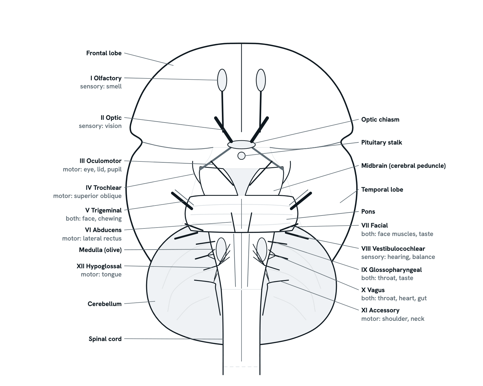
All twelve cranial nerves seen from under the brain, in order from front to back, with each one's function, skull opening and bedside test.
The brain from below, front at the top, simplified and not to scale: the frontal lobes with the olfactory bulbs, the optic nerves meeting at the chiasm, the temporal lobes at the sides, then the midbrain, pons and medulla in front of the cerebellum. The twelve nerves leave in numbered order from front to back; both sides' roots are drawn, each pair labelled once. Press any nerve for its job, skull opening and test.
Also called 12 cranial nerves, Cranial nerves chart, Cranial nerve mnemonic or Cranial nerves and their functions.
Press any part of the diagram, or pick one here, to see what it is and what it does.
Download the diagramChecked against its sources on 5 November 2026.
A tour
A tour of the cranial nerves
01Twelve pairs, numbered from front to back
Twelve pairs of nerves leave the brain and brainstem directly, unlike the spinal nerves, which leave the cord. They are numbered I to XII in the order they attach from front to back, so the number tells you where to look: I and II on the forebrain, III and IV on the midbrain, V to VIII on the pons, IX to XII on the medulla. Three are purely sensory (I, II, VIII), five purely motor (III, IV, VI, XI, XII) and four carry both (V, VII, IX, X).
02I and II: the forebrain
The first two are not ordinary peripheral nerves. The olfactory nerve is the fine filaments that rise from the nose through the cribriform plate into the olfactory bulb; the bulb and tract, part of the brain itself, carry smell back to the temporal lobe. The optic nerve is an outgrowth of the brain, myelinated by oligodendrocytes, not Schwann cells; it brings vision from the eye through the optic canal to the chiasm, where the fibres from the nasal half of each retina cross.
03III and IV: the midbrain
The oculomotor nerve leaves the interpeduncular fossa between the two cerebral peduncles and runs forward to the orbit. The trochlear nerve is the odd one out: it leaves the back of the midbrain, below the inferior colliculus, having crossed to the other side, then winds round the peduncle to appear at its side. Both pass through the cavernous sinus and the superior orbital fissure.
04V to VIII: the pons
The trigeminal nerve, the largest, leaves the side of the pons. Along the lower border of the pons, where it meets the medulla, three more leave: the abducens near the midline, and the facial and vestibulocochlear nerves at the side, in the angle between pons and cerebellum, from which they travel together into the internal acoustic meatus.
05IX to XII: the medulla
Four nerves leave the medulla. The glossopharyngeal, vagus and accessory nerves leave in a row behind the olive and pass together through the jugular foramen; the hypoglossal nerve leaves in front of the olive, between it and the pyramid, and has a canal of its own. The accessory nerve's spinal root comes up from the cervical cord through the foramen magnum to join them.
06Sensory, motor or both: Some Say Marry Money
Some Say Marry Money But My Brother Says Big Brains Matter More gives the type of each nerve in order: S for sensory, M for motor, B for both. So I, II and VIII are sensory; III, IV, VI, XI and XII motor; V, VII, IX and X mixed. Four nerves also carry parasympathetic fibres to glands, the eye and the organs: III, VII, IX and X.
07The names: Oh Oh Oh To Touch And Feel
Oh Oh Oh To Touch And Feel Very Good Velvet, Such Heaven: Olfactory, Optic, Oculomotor, Trochlear, Trigeminal, Abducens, Facial, Vestibulocochlear, Glossopharyngeal, Vagus, Spinal accessory, Hypoglossal. Say it with the picture in front of you, matching each word to a root from front to back, and the order will stay with you.
08III, IV and VI move the eye
Three nerves move the eye. The oculomotor supplies four of its six muscles and the lid and constricts the pupil; the trochlear supplies only the superior oblique; the abducens only the lateral rectus. All three enter the orbit through the superior orbital fissure and are tested together with a finger moved in an H. A third nerve palsy leaves the eye down and out with a drooping lid; a sixth stops it turning outwards; a fourth gives vertical double vision, worst looking down.
09I, II and VIII: the special senses
Smell, sight, and hearing with balance each have a nerve of their own, and all three are purely sensory. They are tested in turn: a familiar scent in each nostril with the eyes closed; the acuity chart, the visual fields by confrontation, the pupils' response to light and the back of the eye; a whisper or rubbed fingers at each ear, then tuning-fork tests to tell conductive from sensorineural hearing loss.
10The vagus, the wanderer
The vagus is the longest cranial nerve and the one that leaves the head and neck: from the jugular foramen it runs down beside the carotid artery into the chest and abdomen. It moves the palate, pharynx and larynx, slows the heart, constricts the airways and drives the gut as far as the splenic flexure, and most of its fibres are sensory, reporting back from these organs. Its recurrent laryngeal branch can be injured in thyroid surgery, leaving the voice hoarse.
11Testing them at the bedside, in order
Clinicians test the nerves in order. I: a scent in each nostril. II: acuity, fields, pupils, fundus. III, IV, VI: follow a finger in an H. V: light touch on forehead, cheek and jaw, clench the teeth, corneal reflex. VII: raise the eyebrows, shut the eyes tight, show the teeth, puff the cheeks. VIII: whisper, tuning forks. IX, X: say ah, watch the palate and uvula, gag reflex, voice. XI: shrug and turn the head against resistance. XII: stick out the tongue.
Parts
What each part of the cranial nerves does
- I Olfactory nerve
- Sensory: smell. Fine fibres from the nose pass through the holes of the cribriform plate of the ethmoid bone and end in the olfactory bulb on the under-surface of the frontal lobe; the tract carries the signal back towards the temporal lobe. A head injury can shear these fibres at the plate and abolish smell (anosmia).
- II Optic nerve
- Sensory: vision. The axons of 1.2 million retinal ganglion cells form each nerve, which enters the skull by the optic canal to meet its fellow at the chiasm. An outgrowth of the brain, myelinated by oligodendrocytes, not Schwann cells. Optic neuritis, often in multiple sclerosis, blurs one eye and hurts on moving it.
- III Oculomotor nerve
- Motor. From the interpeduncular fossa it runs through the cavernous sinus and superior orbital fissure to four of the six eye muscles and the lid's levator, plus parasympathetic fibres to the pupil. A palsy leaves the eye down and out with a drooping lid; a fixed, dilated pupil suggests an aneurysm pressing on it.
- IV Trochlear nerve
- Motor to the superior oblique only. The thinnest cranial nerve, the only one to leave the back of the brainstem; it crosses to the other side, winds round the midbrain and enters the orbit. A palsy gives vertical double vision, worst looking down and in, and a head tilt away from the weak eye.
- V Trigeminal nerve
- Both, and the largest cranial nerve. It leaves the side of the pons with a large sensory and a small motor root and divides at its ganglion into the ophthalmic, maxillary and mandibular nerves. It feels the face, eye, nose, mouth and teeth; V3 moves the chewing muscles. Trigeminal neuralgia is its electric-shock pain.
- VI Abducens nerve
- Motor to the lateral rectus, which turns the eye outwards. It leaves where pons meets medulla, near the midline, runs far forward to the cavernous sinus and orbit. A palsy stops the eye abducting, with horizontal double vision worst looking that way; raised pressure in the skull can damage it.
- VII Facial nerve
- Both. From the pontomedullary junction it enters the internal acoustic meatus and leaves the skull at the stylomastoid foramen for the muscles of facial expression. It also carries taste from the front two-thirds of the tongue and drives the tear and salivary glands. Bell's palsy is its sudden one-sided weakness.
- VIII Vestibulocochlear nerve
- Sensory: hearing and balance. Its cochlear and vestibular parts run with the facial nerve through the internal acoustic meatus. A vestibular schwannoma (acoustic neuroma), a benign tumour of its Schwann cells, grows here, causing one-sided hearing loss, tinnitus and unsteadiness; about one person in 100,000 a year.
- IX Glossopharyngeal nerve
- Both. It leaves the side of the medulla and the skull by the jugular foramen. It carries taste and sensation from the back third of the tongue and pharynx, senses blood pressure at the carotid sinus, drives the parotid gland and moves the stylopharyngeus. Sensory limb of the gag reflex; the vagus is the motor limb.
- X Vagus nerve
- Both; the longest cranial nerve. It leaves the medulla as rootlets behind the olive, passes the jugular foramen and wanders down to the abdomen. It moves palate, pharynx and larynx and supplies the heart, lungs and gut; most of its fibres are sensory. A palsy: hoarse voice, uvula pulled to the healthy side.
- XI Accessory nerve
- Motor to the sternocleidomastoid and trapezius. Most of it arises from the upper cervical cord: its spinal roots climb through the foramen magnum into the skull and leave again, with IX and X, through the jugular foramen. Damage weakens the shrug and turning the head to the other side, and the shoulder droops.
- XII Hypoglossal nerve
- Motor to the tongue muscles. Its rootlets leave the medulla between the pyramid and the olive and gather into the hypoglossal canal. Asked to stick the tongue out, a patient with a damaged nerve sees it deviate towards the weak side, pushed across by the healthy genioglossus; in time that side wastes and twitches.
- Frontal lobes
- The under-surface of the frontal lobes, with the longitudinal fissure between them. The olfactory bulb and tract lie here, between the gyrus rectus beside the midline and the orbital gyri, in the anterior cranial fossa.
- Temporal lobes
- The temporal lobes bulge out at the sides, separated from the frontal lobe in front by the stem of the lateral sulcus. Smell is perceived on their inner surface, in the piriform cortex and amygdala, where the olfactory tract ends.
- Optic chiasm
- Where the two optic nerves meet. Fibres from the nasal half of each retina, which see the outer half of the visual field, cross here to the other side; those from the temporal half stay on their own side. A pituitary tumour pressing on the chiasm cuts off both outer fields: bitemporal hemianopia.
- Pituitary stalk
- The cut stalk, or infundibulum, by which the pituitary gland hangs from the base of the brain, just behind the optic chiasm. This closeness is why a pituitary tumour growing upwards presses on the chiasm and narrows the visual fields.
- Midbrain
- The top of the brainstem, mostly hidden between the temporal lobes. From below, its two cerebral peduncles run up from the pons into the hemispheres with the interpeduncular fossa between them. The oculomotor nerve (III) leaves from the fossa; the trochlear (IV) leaves the back of the midbrain and winds round its side.
- Pons
- The bulging middle part of the brainstem. The trigeminal nerve (V) leaves its side, and the abducens (VI), facial (VII) and vestibulocochlear (VIII) nerves leave along its lower border, where it meets the medulla.
- Medulla oblongata
- The lowest part of the brainstem, which continues down as the spinal cord. Beside the midline run the pyramids, and lateral to them the oval olives. The hypoglossal nerve (XII) leaves between pyramid and olive; the glossopharyngeal (IX), vagus (X) and accessory (XI) nerves leave in a row behind the olive.
- Cerebellum
- Behind the brainstem, its two hemispheres show below the back of the cerebrum. In the angle between it and the pons, the cerebellopontine angle, the facial and vestibulocochlear nerves leave for the internal acoustic meatus, and a vestibular schwannoma grows here and in that canal.
- Spinal cord
- The medulla becomes the spinal cord at the foramen magnum. The spinal root of the accessory nerve (XI) arises from the cord's upper cervical segments and climbs up beside it through the foramen magnum into the skull.
As a table
The cranial nerves as a table
The twelve cranial nerves in order, with the type of fibres each carries, its main functions, the opening through which it leaves the skull and a quick bedside test.
I Olfactory
- Type
- Sensory
- Main functions
- Smell
- Leaves the skull through
- Cribriform plate of the ethmoid bone
- Quick bedside test
- Identify a familiar scent in each nostril, eyes closed
II Optic
- Type
- Sensory
- Main functions
- Vision; sensory limb of the pupillary light reflex
- Leaves the skull through
- Optic canal
- Quick bedside test
- Acuity chart, visual fields by confrontation, pupil reactions, fundoscopy
III Oculomotor
- Type
- Motor (with parasympathetic fibres)
- Main functions
- Superior, inferior and medial rectus, inferior oblique and levator palpebrae; constricts the pupil and focuses the lens
- Leaves the skull through
- Superior orbital fissure
- Quick bedside test
- Follow a finger in an H; look for a drooping lid, a dilated pupil, an eye turned down and out
IV Trochlear
- Type
- Motor
- Main functions
- Superior oblique: turns the eye down and rotates it inwards (intorsion)
- Leaves the skull through
- Superior orbital fissure
- Quick bedside test
- Look down and in; vertical double vision and a head tilt away from the weak eye
V Trigeminal
- Type
- Both
- Main functions
- Sensation from the face, scalp, eye, nose, mouth and teeth; the chewing muscles (V3)
- Leaves the skull through
- Superior orbital fissure (V1), foramen rotundum (V2), foramen ovale (V3)
- Quick bedside test
- Light touch in the three divisions, the corneal reflex, clench the teeth and feel the masseters
VI Abducens
- Type
- Motor
- Main functions
- Lateral rectus: turns the eye outwards
- Leaves the skull through
- Superior orbital fissure
- Quick bedside test
- Look to each side; the weak eye fails to turn outwards
VII Facial
- Type
- Both
- Main functions
- Muscles of facial expression and the stapedius; taste from the front two-thirds of the tongue; lacrimal and salivary glands (not the parotid)
- Leaves the skull through
- Internal acoustic meatus, then the stylomastoid foramen
- Quick bedside test
- Raise the eyebrows, shut the eyes tight, show the teeth, puff out the cheeks
VIII Vestibulocochlear
- Type
- Sensory
- Main functions
- Hearing (cochlear part) and balance (vestibular part)
- Leaves the skull through
- Internal acoustic meatus
- Quick bedside test
- Whispered words or rubbed fingers at each ear; Rinne and Weber tuning-fork tests
IX Glossopharyngeal
- Type
- Both
- Main functions
- Taste and sensation from the back third of the tongue and the pharynx; carotid sinus and body; parotid gland; stylopharyngeus
- Leaves the skull through
- Jugular foramen
- Quick bedside test
- Gag reflex (its sensory limb) and swallowing, tested together with X
X Vagus
- Type
- Both
- Main functions
- Palate, pharynx and larynx; parasympathetic supply to the heart, lungs and gut as far as the splenic flexure; sensation from the organs
- Leaves the skull through
- Jugular foramen
- Quick bedside test
- Say ah: the palate rises evenly and the uvula stays central; voice, cough, gag reflex (its motor limb)
XI Accessory
- Type
- Motor
- Main functions
- Sternocleidomastoid and trapezius
- Leaves the skull through
- Jugular foramen (its spinal root enters through the foramen magnum)
- Quick bedside test
- Shrug the shoulders and turn the head against resistance
XII Hypoglossal
- Type
- Motor
- Main functions
- Muscles of the tongue
- Leaves the skull through
- Hypoglossal canal
- Quick bedside test
- Stick out the tongue: it deviates towards the weak side; push it into each cheek
The numbers follow the order in which the nerves attach to the brain from front to back. The bedside tests are those of the StatPearls chapter Cranial Nerve Testing; IX and X are tested together because they share the palate, pharynx and gag reflex.
Dictionary
The terms to know
Practise it
The quizzes and cards that fit
- Neuroscience quizzes
- Reflex Arc15 questions
- Spinal Cord15 questions
- Cognitive Neuroscience: Language & Lateralization15 questions
- Human Anatomy quizzes
- Shoulder and Rotator Cuff15 questions
- Knee Joint15 questions
- Arteries and Veins15 questions
- Medicine quizzes
- Labor and Delivery15 questions
- IV Fluids15 questions
- Sepsis15 questions
- Neuroscience flashcards
The subject's terms as a ready-made deck, free to flip through.
Next to it
More diagrams
Sources
Where the facts come from
- StatPearls: Neuroanatomy, Cranial Nerve (NCBI Bookshelf)
- StatPearls: Cranial Nerve Testing (NCBI Bookshelf)
- StatPearls: Anatomy, Head and Neck, Skull Foramen (NCBI Bookshelf)
- StatPearls: Neuroanatomy, Cranial Nerve 2 (Optic) (NCBI Bookshelf)
- StatPearls: Optic Neuritis (NCBI Bookshelf)
- StatPearls: Neuroanatomy, Cranial Nerve 3 (Oculomotor) (NCBI Bookshelf)
- StatPearls: Trochlear Nerve Palsy (NCBI Bookshelf)
- StatPearls: Neuroanatomy, Cranial Nerve 5 (Trigeminal) (NCBI Bookshelf)
- StatPearls: Neuroanatomy, Cranial Nerve 10 (Vagus Nerve) (NCBI Bookshelf)
- Romano and others, Imaging of cranial nerves: a pictorial overview, Insights into Imaging 2019 (PMC)
- Libreros-Jiménez and others, On the Cranial Nerves, NeuroSci 2024 (PMC)
- Finsterer and Grisold, Disorders of the lower cranial nerves, Journal of Neurosciences in Rural Practice 2015 (PMC)
- Patros and others, The physiology, anatomy and stimulation of the vagus nerve in epilepsy, Journal of Physiology 2025 (PMC)
- Singh and Deshmukh, Bell's Palsy: A Review, Cureus 2022 (PMC)
- Xu and others, Trigeminal neuralgia: from clinical challenges to molecular insights, Journal of Headache and Pain 2026 (PMC)
- Oculomotor Nerve Palsy: Etiologies, Symptoms and Diagnosis, a systematic review with meta-analysis, Diagnostics 2026 (PMC)
- Alexandrou and others, Trochlear Nerve Palsy: A Systematic Review of Etiologies and Diagnostic Insights, Diagnostics 2025 (PMC)
- NIDCD: Vestibular Schwannoma (Acoustic Neuroma) and Neurofibromatosis
Drawn and written by biofideX from these sources.